
Cardiology
Category: Abstract Submission
Cardiology I

Howard H. Lei, Ph.D.
Senior Data Scientist
CHOC Children's Hospital of Orange County
Los Angeles, California, United States
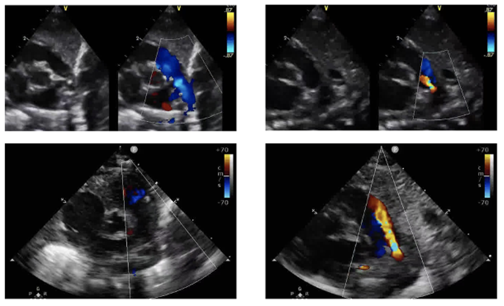 This figure shows four sample echocardiogram video frames. The two images on the right of Figure 3 show examples of a PDA, while the images on the left do not contain a PDA. The PDA is indicated by the red-colored flow pattern during the diastolic phase of the cardiac cycle. The two images in the top row of Figure 3 are color-compare scans. They contain two scans side-by-side, one with color Doppler flow mapping and one without color Doppler. The two scans in the bottom row contain non-color compare scans, with just a single scan showing colored Doppler flow mapping. The goal of this work is to indiscriminately handle the color-compare and non-color compare scans, as both are encountered in real-world scenarios.
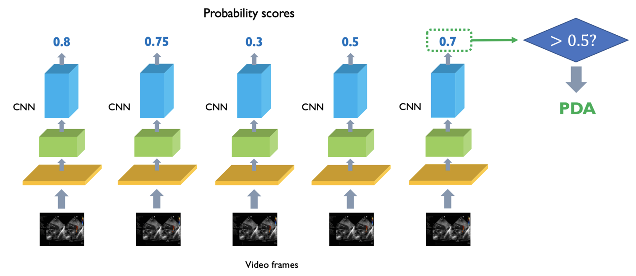 Processing of video clip frames as independent images using the MobileNet-V2 CNN. The CNN generates a probably score for each video frame, indicating the probability of whether the video frame contains a PDA. If the probability exceeds 50%, the video frame is classified as containing a PDA.