Pediatric Neurology
Category: Abstract Submission
Neurology 1: Clinical Pediatric & Neonatal Neurology
548 - The promising role of brain magnetic resonance elastography in evaluation of pediatric brain tumors
Friday, April 22, 2022
6:15 PM - 8:45 PM US MT
Poster Number: 548
Publication Number: 548.139
Publication Number: 548.139
Abdulhafeez Khair, Nemours Children's Hospital, Wilmington, DE, United States; Grace Mcllvain, university of delaware, Wilmington, DE, United States; Curtis Johnson, University of Delaware, Newark, DE, United States; Vinay Kandula, Sidney Kimmel Medical College at Thomas Jefferson University, Wallingford, PA, United States; Lauren W. Averill, Sidney Kimmel Medical College at Thomas Jefferson University, Wilmington, DE, United States; Gurcharanjeet Kaur, Nemours Children's Hospital, Wilmington, DE, United States; Andrew Walter, Nemours Children's Hospital, Wilmington, DE, United States; Rahul Nikam, Nemours Children's Hospital, Wilmington, DE, United States

Hafeez Khair, MD, MHPE, MRCPCH-UK
Fellow
Nemours Children's Hospital
Wilmington, Delaware, United States
Presenting Author(s)
Background: Medical imaging okays an invaluable role in diagnosis, treatment, & monitoring of pediatric brain tumors, but is critically met with low specificity and lack of histological correlation. A promising imaging modality for probing brain structure in both health and disease stats is magnetic resonance elastography (MRE), an emerging technique for evaluation of viscoelastic mechanical properties of human tissues as a quantitative non-invasive palpation. Brain MRE assesses how tissue elements act and interact when mechanically forced and provide critical information about distribution and organization of neurons, glial cells, & extracellular matrix.
Objective: To Compare the stiffness of pediatric brain tumors with stiffness of uninvolved contralateral white matter and to try determining the relationship of tumor stiffness with tumor grade.
Design/Methods: We prospectively involved 17 subjects with working diagnosis of pediatric brain tumors, with a minimum tumor dimension of 15 mm from July 2020 to December 2021). All studies were performed using a GE Signa 2T PET/MR scanner. Single-shot echo-planar imaging (EPI) sequence was used for fast, motion-robust imaging of MRE displacement data with whole-brain coverage and 2.5 mm isotropic spatial resolution. The MRE sequence was synchronized with external vibration at 50 Hz delivered via a pneumatic actuator and soft pillow driver. The reported stiffness (μ) and damping ratio (ɛ) were then calculated.
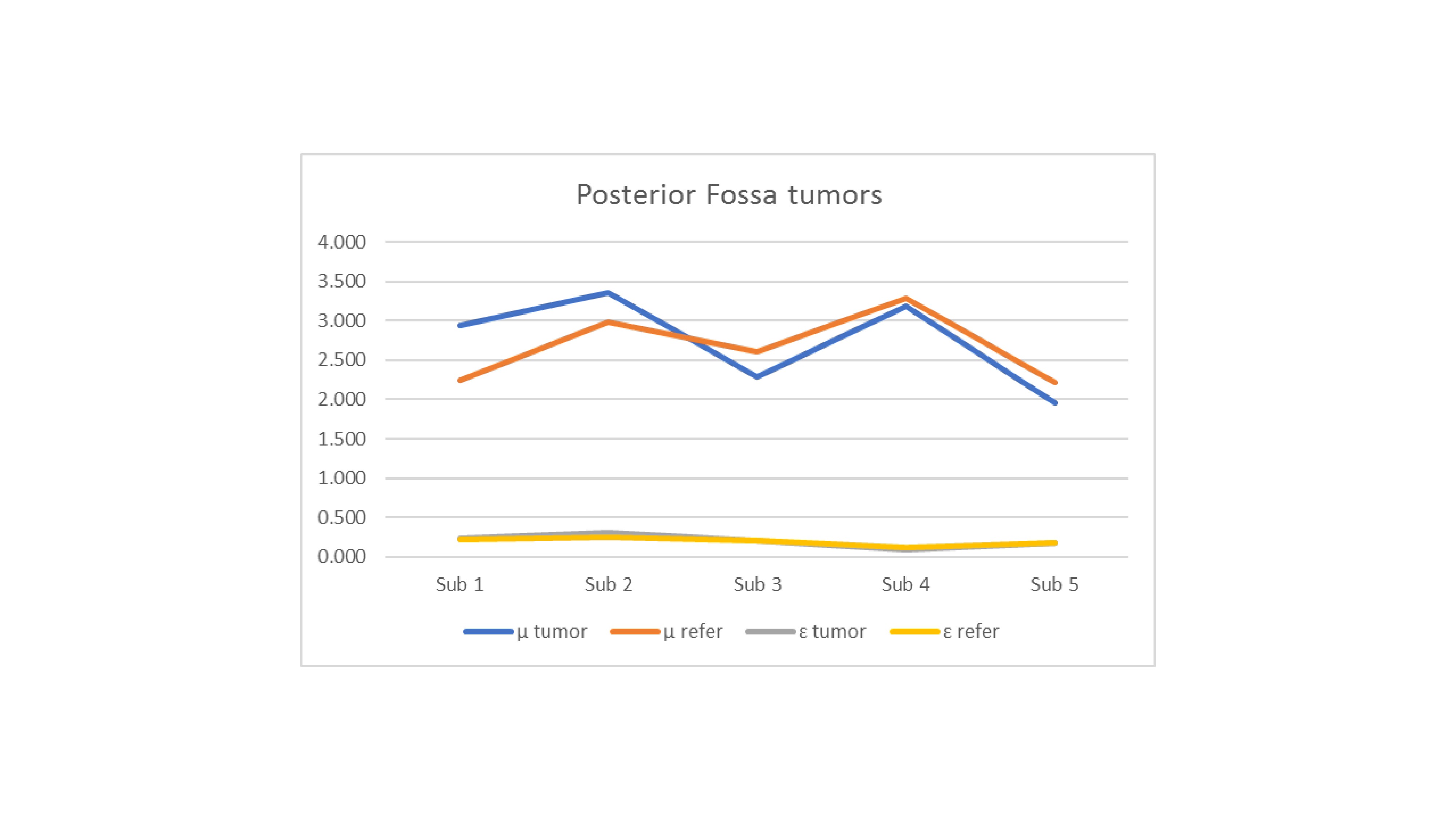
Results: 12 subjects had supratentorial tumors whereas 5 subjects had cerebellar or brainstem tumors. Supratentorial tumors were significantly less stiff, with mean stiffness of 2.487 ± 0.293, reference mean of 2.976 ± 0.188 and P value of 0.0117. Interestingly, cerebellar and brainstem tumors were slightly stiffer with mean stiffness of 2.746 ± 0.469 & reference mean of 2.669 ± 0.363 but the difference was not statistically significant with P value of 0.805. Damping ratio means were 0.253 ± 0.096 in the supratentorial group, with reference mean of 0.233 ± 0.015 and P value of 0.6805, & 0.205 ± 0.064, with reference mean of 0.195 ± 0.04 & P value of 0.8058. in the infratentorial group.Conclusion(s): Brain MRE is a relatively new, rapidly evolving, special sequence of MRI-based technology that relies on mapping the elastic properties of soft body tissues. In our novel pilot study utilizing brain MRE, supratentorial tumors had significantly lower stiffness, whereas cerebellar tumors had slightly higher stiffness in reference to healthy looking tissues. Pediatric brain MRE can have a very promising role in improving the diagnostic precision of pediatric brain tumors.
Khair CV-2021.pdf
Figure 2

