Cardiology
Category: Abstract Submission
Cardiology II
89 - Cardiovascular Magnetic Resonance in Children with Multisystem Inflammatory Syndrome in Children (MIS-C) Associated with COVID-19 – Institutional Protocol based Medium-term Follow-up study
Sunday, April 24, 2022
3:30 PM - 6:00 PM US MT
Poster Number: 89
Publication Number: 89.303
Publication Number: 89.303
Abhishek Chakraborty, University of Tennessee Health Science Center College of Medicine, Memphis, TN, United States; Ranjit Philip, University of Tennessee Health Science Center College of Medicine, Memphis, TN, United States; Michelle Santoso, University of Tennessee Health Science Center College of Medicine, Memphis, TN, United States; Ronak Naik, University of Tennessee Health Science Center College of Medicine, Memphis, TN, United States; Anthony Merlocco, Le Bonheur Children's Hospital, Memphis, TN, United States; Jason N. Johnson, Le Bonheur Children's Hospital, Memphis, TN, United States

Abhishek Chakraborty, MD, FACC
Assistant Professor, Pediatric Cardiology
University of Tennessee Health Sciences
Memphis, Tennessee, United States
Presenting Author(s)
Background: Multisystem inflammatory syndrome in children (MIS-C) secondary to COVID-19 infection has been a major cause of cardiovascular morbidity in children and adolescents. Although there is growing pool of data to suggest systolic ventricular dysfunction function and coronary artery abnormalities almost always recover within 3-6 months, subtle abnormalities in diastolic function and evidence of myocardial injury persists.
Objective:
Investigate medium term cardiovascular outcomes of patients with MIS-C using CMR
Design/Methods:
This is a single center retrospective study of patients aged less than 21-years, diagnosed with MIS-C who received an outpatient CMR (cardiovascular magnetic resonance), around 6 months after discharge. Cardiac biomarkers, including Troponin I, brain natriuretic peptide (BNP), and echocardiographic data were collected at peak of illness during admission, at 6-weeks and 6-months after discharge. CMR was done in patients with significant troponin leak or depressed LVEF. All CMR performed on a GE Signa HDxt 1.5 Tesla magnet with a myocarditis protocol (standard cine steady state free precession, T2 weighted short axis stack, early gadolinium enhancement short axis stack, late gadolinium enhancement short axis stack and long axis images (segmented GRE post contrast)). Diagnosis of myocarditis was determined by the original Lake Louise Criteria.
Results:
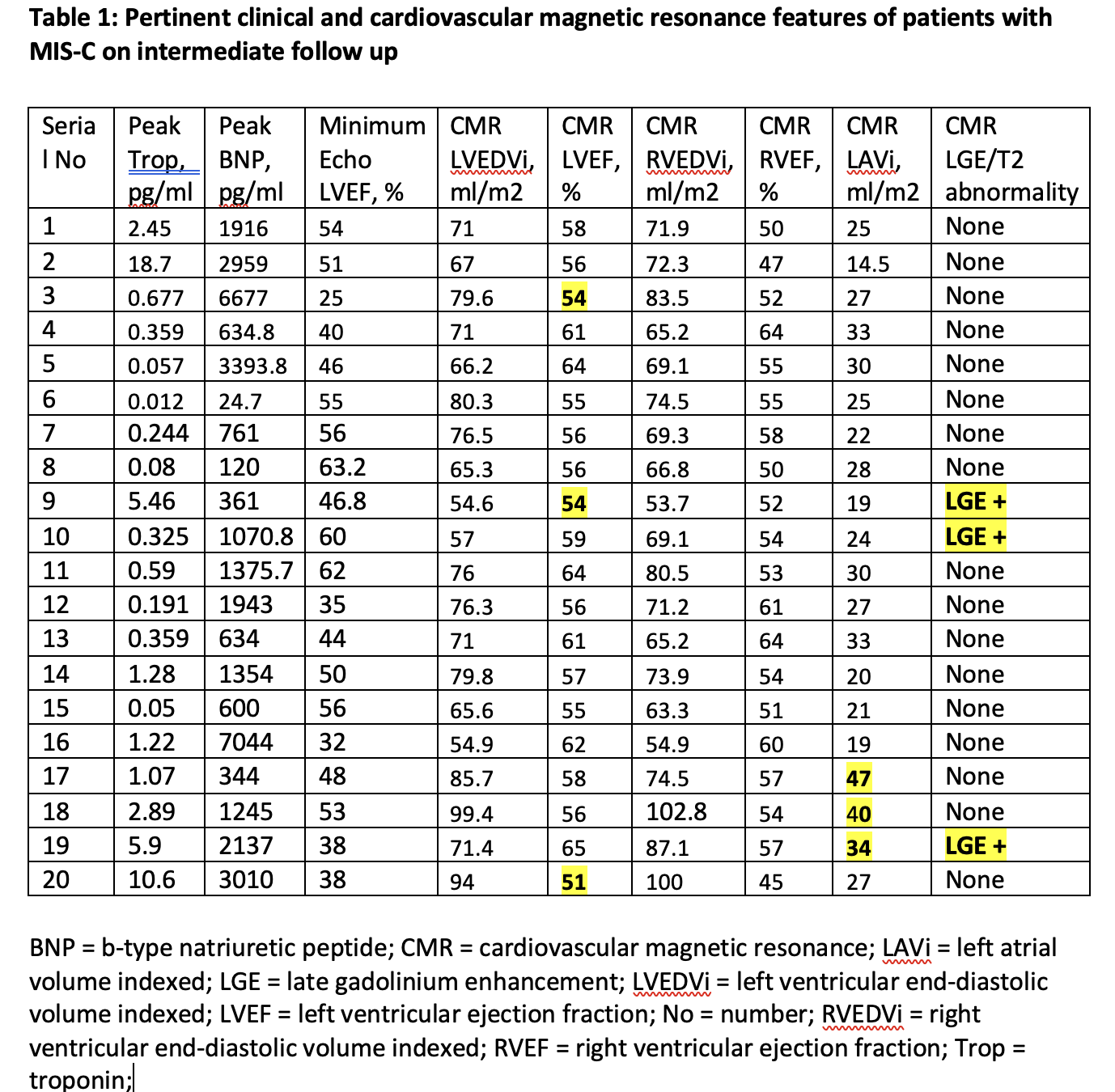
There were 20 patients with a median age of 11 years, (IQR 8-13 years), who underwent CMR at median follow-up duration of 6 months (IQR 5-7 months). At the peak of illness during admission there were 95% patients with abnormal Troponin I and BNP. By echocardiogram 70% had left ventricular systolic dysfunction including 15% with severe dysfunction. There was complete recovery of left ventricular systolic function at 6 weeks. At the peak of illness, there were 15% had prominent coronaries and 10% had coronary ectasia, which all resolved by 6 months. By CMR, there were 5 patients (25%) with abnormal left atrial volume, 1 patient (5%) with an abnormal indexed left ventricular end-diastolic volume, and 3 patients (15%) with abnormal LVEF. None of the patients had any coronary abnormalities. There was no evidence of myocardial edema in T2 weighted image sequence in any of the patients. There were 3 patients with persistent late gadolinium enhancement on CMR.
Conclusion(s):
MIS-C has a heterogenous cardiovascular manifestation and a spectrum of underlying pathophysiology. Follow-up CMR is a useful tool in diagnosing subtle myocardial abnormalities and guide necessity for future follow-up.
Late CMR findings in MIS-C Figure 1. Cardiovascular magnetic resonance short axis late gadolinium enhancement in three separate patients. A. Patient 1. Apical segment with epicardial enhancement in the lateral wall (arrow). B. Patient 2. Mid segment with mid myocardial enhancement in the anteroseptal wall (arrow). C. Patient 3. Mid segment with epicardial enhancement in the anterior wall (arrow).
Pertinent clinical and cardiovascular magnetic resonance features of patients with MIS-C on intermediate follow up
Objective:
Investigate medium term cardiovascular outcomes of patients with MIS-C using CMR
Design/Methods:
This is a single center retrospective study of patients aged less than 21-years, diagnosed with MIS-C who received an outpatient CMR (cardiovascular magnetic resonance), around 6 months after discharge. Cardiac biomarkers, including Troponin I, brain natriuretic peptide (BNP), and echocardiographic data were collected at peak of illness during admission, at 6-weeks and 6-months after discharge. CMR was done in patients with significant troponin leak or depressed LVEF. All CMR performed on a GE Signa HDxt 1.5 Tesla magnet with a myocarditis protocol (standard cine steady state free precession, T2 weighted short axis stack, early gadolinium enhancement short axis stack, late gadolinium enhancement short axis stack and long axis images (segmented GRE post contrast)). Diagnosis of myocarditis was determined by the original Lake Louise Criteria.
Results:
There were 20 patients with a median age of 11 years, (IQR 8-13 years), who underwent CMR at median follow-up duration of 6 months (IQR 5-7 months). At the peak of illness during admission there were 95% patients with abnormal Troponin I and BNP. By echocardiogram 70% had left ventricular systolic dysfunction including 15% with severe dysfunction. There was complete recovery of left ventricular systolic function at 6 weeks. At the peak of illness, there were 15% had prominent coronaries and 10% had coronary ectasia, which all resolved by 6 months. By CMR, there were 5 patients (25%) with abnormal left atrial volume, 1 patient (5%) with an abnormal indexed left ventricular end-diastolic volume, and 3 patients (15%) with abnormal LVEF. None of the patients had any coronary abnormalities. There was no evidence of myocardial edema in T2 weighted image sequence in any of the patients. There were 3 patients with persistent late gadolinium enhancement on CMR.
Conclusion(s):
MIS-C has a heterogenous cardiovascular manifestation and a spectrum of underlying pathophysiology. Follow-up CMR is a useful tool in diagnosing subtle myocardial abnormalities and guide necessity for future follow-up.
Late CMR findings in MIS-C
 Figure 1. Cardiovascular magnetic resonance short axis late gadolinium enhancement in three separate patients. A. Patient 1. Apical segment with epicardial enhancement in the lateral wall (arrow). B. Patient 2. Mid segment with mid myocardial enhancement in the anteroseptal wall (arrow). C. Patient 3. Mid segment with epicardial enhancement in the anterior wall (arrow).
Pertinent clinical and cardiovascular magnetic resonance features of patients with MIS-C on intermediate follow up

