Neonatal Neurology: Translational
Category: Abstract Submission
Neurology 5: Neonatal Neurology Term Clinical
484 - Prenatal COVID exposure affects neonatal brain development
Sunday, April 24, 2022
3:30 PM - 6:00 PM US MT
Poster Number: 484
Publication Number: 484.343
Publication Number: 484.343
Nickie Andescavage, Children's National Health System, Washington, DC, United States; Yuan-Chiao Lu, Children's National Hospital, Washington, DC, United States; Catherine Lopez, Children's National Hospital, Washington, DC, United States; Kushal Kapse, Developing Brain Institute, Washington, DC, United States; Catherine Lopez, Children's National Health System, Washington, D.C., DC, United States; Jessica Quistorff, Children's National Health System, Washington, DC, United States; Nicole Andersen, Children's National Health System, Washington, DC, United States; Gilbert Vezina, Children's National Health System, Washington, DC, United States; Diedtra Henderson, Children's National Health System, Washington, DC, United States; David Wessel, Children's National Hospital, Washington, DC, United States; Scott Barnett, Children's National Health System, Arlington, VA, United States; Adre du Plessis, George Washington University School of Medicine and Health Sciences, Washington, DC, United States; Catherine Limperopoulos, Children's National Health System, Washington, DC, United States; Yao Wu, Children's National Hospital, Washington, DC, United States

Nickie Andescavage, MD
Associate Professor
Children's National Health System
Washington, District of Columbia, United States
Presenting Author(s)
Background: The novel virus, SARS-COV-2 was declared a global pandemic in 2020 and continues unabated nearly two years later. Prenatal viral exposures may influence early brain development and predispose offspring to neuropsychiatric conditions later in life. While vertical transmission is rare, secondary effects of maternal COVID-19 infection in pregnancy remain largely unknown.
Objective: To determine the association between maternal COVID-19 infection in pregnancy and offspring neurodevelopment
Design/Methods: Prospectively enrolled underwent brain MRI at term-equivalent age, and global and tissue-specific brain volumes (cortical grey matter, white matter, deep grey matter (DGM), cerebellum, brainstem and total brain) were measured. Global and regional cortical features such as surface area, gyrification index, and sulcal depth were also measured. Comparisons of these measurements were made to a pre-pandemic cohort of healthy control newborns. Group comparisons were adjusted for gestational age at MRI, infant race, and infant sex with between group post-hoc tests; data presented as adjusted means±SE.
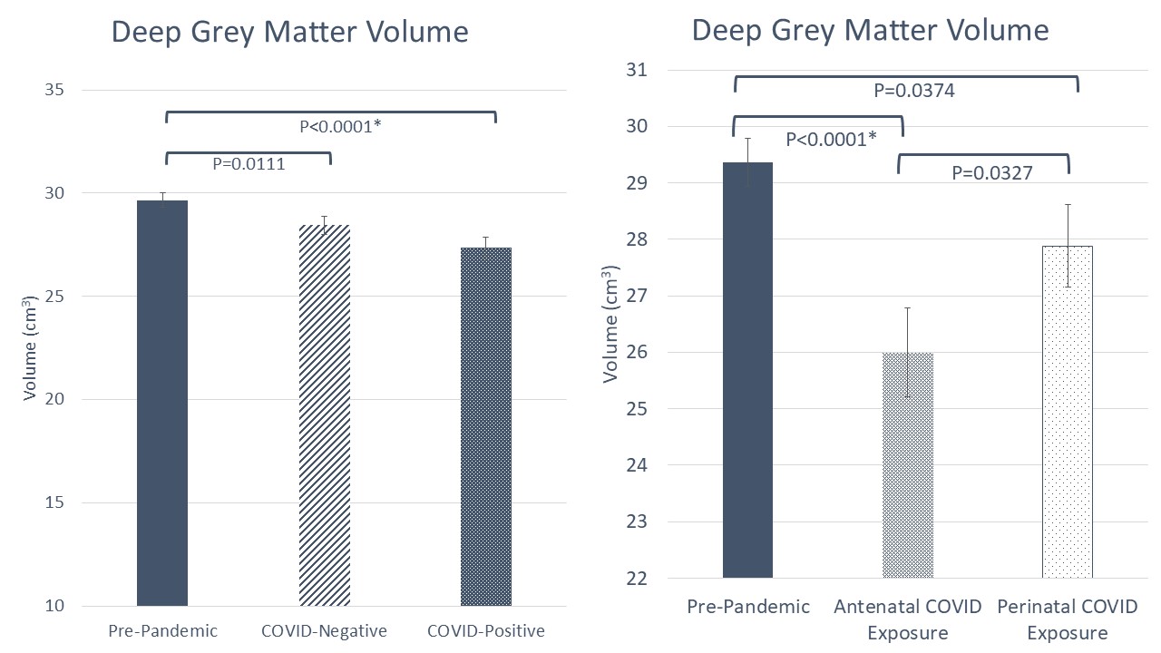
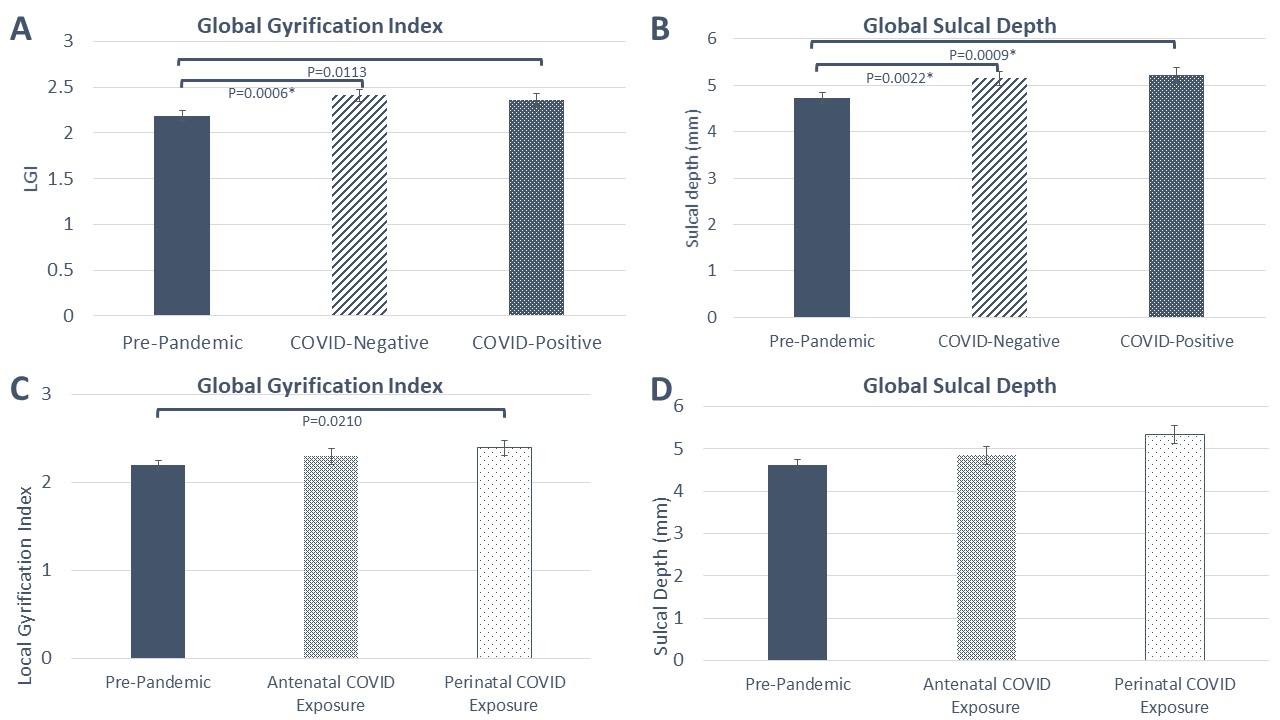
Results: We evaluated 210 infants (45 infants of COVID-19 unexposed mothers, 47 infants of COVID-19 positive mothers, 108 pre-pandemic controls). We found reduced DGM in infants of COVID-19 positive mothers (27.35 ± 0.53 vs 29.65 ± 0.35 (pre-pandemic controls, p< 0.01), with the most significant decrease in those exposed prior to delivery (Fig 1). We also observed an increase in gyrification and sulcal depth in infants of both COVID-19 unexposed and COVID-19 positive mothers compared to pre-pandemic controls (Table 1, Fig 2). For infants born to COVID-19 positive mothers, antenatal exposure was significant for decreased surface area of the occipital lobe compared to those with perinatal exposure (90.69± 5.68 vs. 105.61 ± 5.10, p=0.02), decreased sulcal depth of the parietal (6.48 ± 0.26 vs. 7.07 ± 0.23, p=0.03) and temporal lobes (5.13 ± 0.19 vs. 5.61 ± 0.17, p=0.02)Conclusion(s): Despite the low intrauterine transmission rates of COVID-19, we report for the first time significant differences of regional neonatal brain volumes and cerebral cortical maturation in infants born to COVID-19 positive mothers. These changes may be mediated by secondary factors of maternal exposure, as well as non-viral stressors associated with the pandemic. Ongoing neurodevelopmental follow-up to determine the functional impact of these findings is currently underway.
Figure 1 Deep grey matter volumes in pre-pandemic neonates (solid bar), compared to infants of COVID-19 unexposed and COVID-19 positive mothers; * denotes q < 0.05 (panel A); Of those infants with COVID-19 positive mothers, antenatal exposure showed the greatest reduction in volumes (panel B).
Figure 2 Global cerebral gyrification indices (panels A/C) and sulcal depths (panels B/D) of pre-pandemic healthy infants (solid bar) compared to infants of COVID-19 unexposed and COVID-19 positive mothers (panels A/B). COVID-19 exposed infants were further evaluated based on antenatal and perinatal exposures (panels C/D). * denotes q < 0.05
Objective: To determine the association between maternal COVID-19 infection in pregnancy and offspring neurodevelopment
Design/Methods: Prospectively enrolled underwent brain MRI at term-equivalent age, and global and tissue-specific brain volumes (cortical grey matter, white matter, deep grey matter (DGM), cerebellum, brainstem and total brain) were measured. Global and regional cortical features such as surface area, gyrification index, and sulcal depth were also measured. Comparisons of these measurements were made to a pre-pandemic cohort of healthy control newborns. Group comparisons were adjusted for gestational age at MRI, infant race, and infant sex with between group post-hoc tests; data presented as adjusted means±SE.
Results: We evaluated 210 infants (45 infants of COVID-19 unexposed mothers, 47 infants of COVID-19 positive mothers, 108 pre-pandemic controls). We found reduced DGM in infants of COVID-19 positive mothers (27.35 ± 0.53 vs 29.65 ± 0.35 (pre-pandemic controls, p< 0.01), with the most significant decrease in those exposed prior to delivery (Fig 1). We also observed an increase in gyrification and sulcal depth in infants of both COVID-19 unexposed and COVID-19 positive mothers compared to pre-pandemic controls (Table 1, Fig 2). For infants born to COVID-19 positive mothers, antenatal exposure was significant for decreased surface area of the occipital lobe compared to those with perinatal exposure (90.69± 5.68 vs. 105.61 ± 5.10, p=0.02), decreased sulcal depth of the parietal (6.48 ± 0.26 vs. 7.07 ± 0.23, p=0.03) and temporal lobes (5.13 ± 0.19 vs. 5.61 ± 0.17, p=0.02)Conclusion(s): Despite the low intrauterine transmission rates of COVID-19, we report for the first time significant differences of regional neonatal brain volumes and cerebral cortical maturation in infants born to COVID-19 positive mothers. These changes may be mediated by secondary factors of maternal exposure, as well as non-viral stressors associated with the pandemic. Ongoing neurodevelopmental follow-up to determine the functional impact of these findings is currently underway.
Figure 1
 Deep grey matter volumes in pre-pandemic neonates (solid bar), compared to infants of COVID-19 unexposed and COVID-19 positive mothers; * denotes q < 0.05 (panel A); Of those infants with COVID-19 positive mothers, antenatal exposure showed the greatest reduction in volumes (panel B).
Figure 2
 Global cerebral gyrification indices (panels A/C) and sulcal depths (panels B/D) of pre-pandemic healthy infants (solid bar) compared to infants of COVID-19 unexposed and COVID-19 positive mothers (panels A/B). COVID-19 exposed infants were further evaluated based on antenatal and perinatal exposures (panels C/D). * denotes q < 0.05