Developmental Biology/Cardiac & Pulmonary Development
Category: Abstract Submission
Developmental Biology/Cardiac & Pulmonary Development I
164 - The Role of Estrogen Receptor Alpha During Lung Development Under Hypoxic Conditions in a Rat Model
Sunday, April 24, 2022
3:30 PM - 6:00 PM US MT
Poster Number: 164
Publication Number: 164.311
Publication Number: 164.311
Nicholas T. Severyn, Indiana University, Indianapolis, IN, United States; Tim Lahm, National Jewish Health, Denver, CO, United States; Robert Tepper, Riley Hospital for Children at Indiana University Health, Indianapolis, IN, United States

Nicholas T. Severyn, DO
Neonatal-Perinatal Fellow
Indiana University
Indianapolis, Indiana, United States
Presenting Author(s)
Background: Humans living at high-altitude have adapted their physiology to survive in this low-oxygen environment by increasing pulmonary vascular and alveolar growth allowing for greater gas exchanging ability. RNA sequencing data from a novel murine model that mimics the human phenotypical response to chronic hypoxia suggest estrogen signaling via estrogen receptor alpha (ERα) is a key mediator in this adaptation.
Objective: We sought to delineate the mechanistic role ERα contributes to this process by exposing novel homozygous loss-of-function ERα mutant rats to a novel murine model of human high-altitude exposure.
Design/Methods: Using rats maintained at normoxia as controls, loss-of-function ERα mutant (ERαMut) rats were exposed to hypoxia (15% FiO2) starting at conception and continued postnatally until 6 weeks of age at which time in vivo and in vitro studies were conducted. (Figure 1) A factorial ANOVA taking into consideration body weight and all three main effects (environment, genotype, and animal sex) was utilized to compare each group for each outcome measured as well, and a p-value of 0.05 was used as significance threshold.
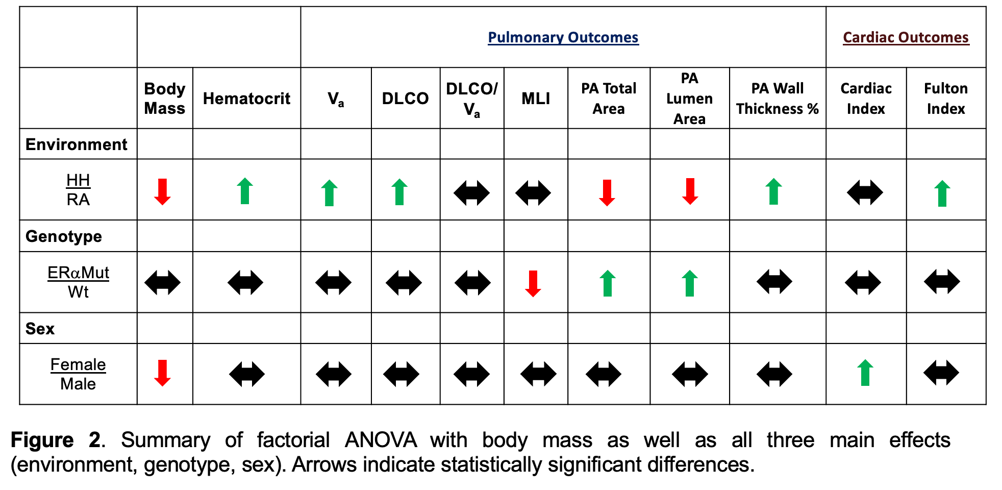
Results: Both wild type (WT) and ERαMut animals born and raised in hypoxia had significantly lower body mass and higher hematocrits, total alveolar volumes (Va), diffusion capacities of carbon monoxide, pulmonary arteriole (PA) wall thickness, and Fulton indices than normoxia WT and ERαMut animals. (Figure 2) No major physiologic differences were seen between WT and ERαMut animals at either exposure, but we did observe histologic differences with ERαMut animals having a smaller mean linear intercept (MLI) and increased PA total and lumen areas. (Figure 2)
Trends for sex differences were noted between males and females, but most did not reach statistical significance. Hypoxia tended to cause less of an increase in Va and PA thickness in females than males, but females in hypoxia tended to have a larger increase in Fulton index. ERαMut males tended to have smaller alveolar volumes and lower PA wall thickness than ERαMut females, but larger PAs (increased PA total area and lumen).
Conclusion(s): At the time point and degree of hypoxia studied, ERα does not seem to have a pivotal role in the cardiopulmonary physiologic adaptation to chronic hypoxia. Given the histologic and potential sex and genotype differences seen, further research into cell-specific effects of ERα during hypoxia-induced cardiopulmonary adaptation is warranted.
Severyn CVSeveryn_CV_Nov2021.pdf
Figure 2 Summary of factorial ANOVA with body mass as well as all three main effects (environment, genotype, sex). Arrows indicate statistically significant differences.
Objective: We sought to delineate the mechanistic role ERα contributes to this process by exposing novel homozygous loss-of-function ERα mutant rats to a novel murine model of human high-altitude exposure.
Design/Methods: Using rats maintained at normoxia as controls, loss-of-function ERα mutant (ERαMut) rats were exposed to hypoxia (15% FiO2) starting at conception and continued postnatally until 6 weeks of age at which time in vivo and in vitro studies were conducted. (Figure 1) A factorial ANOVA taking into consideration body weight and all three main effects (environment, genotype, and animal sex) was utilized to compare each group for each outcome measured as well, and a p-value of 0.05 was used as significance threshold.
Results: Both wild type (WT) and ERαMut animals born and raised in hypoxia had significantly lower body mass and higher hematocrits, total alveolar volumes (Va), diffusion capacities of carbon monoxide, pulmonary arteriole (PA) wall thickness, and Fulton indices than normoxia WT and ERαMut animals. (Figure 2) No major physiologic differences were seen between WT and ERαMut animals at either exposure, but we did observe histologic differences with ERαMut animals having a smaller mean linear intercept (MLI) and increased PA total and lumen areas. (Figure 2)
Trends for sex differences were noted between males and females, but most did not reach statistical significance. Hypoxia tended to cause less of an increase in Va and PA thickness in females than males, but females in hypoxia tended to have a larger increase in Fulton index. ERαMut males tended to have smaller alveolar volumes and lower PA wall thickness than ERαMut females, but larger PAs (increased PA total area and lumen).
Conclusion(s): At the time point and degree of hypoxia studied, ERα does not seem to have a pivotal role in the cardiopulmonary physiologic adaptation to chronic hypoxia. Given the histologic and potential sex and genotype differences seen, further research into cell-specific effects of ERα during hypoxia-induced cardiopulmonary adaptation is warranted.
Severyn CVSeveryn_CV_Nov2021.pdf
Figure 2
 Summary of factorial ANOVA with body mass as well as all three main effects (environment, genotype, sex). Arrows indicate statistically significant differences.