Back
Pediatric Neurology
Category: Abstract Submission
Neurology 7: Neonatal Neurology Term Imaging
347 - Correlation between Argenta classification and cranial asymmetry values in deformational plagiocephaly
Monday, April 25, 2022
3:30 PM – 6:00 PM US MT
Poster Number: 347
Publication Number: 347.443
Publication Number: 347.443
Nobuhiko Nagano, Department of Pediatrics and Child Health, Nihon University School of Medicine, Tokyo, Tokyo, Japan; Takanori Noto, Nihon University Itabashi Hospital, nerima hikawadai 3-40-6, Tokyo, Japan; Risa Kato, Department of Pediatrics and Child Health, Nihon University School of Medicine, Itabashi-ku, Tokyo, Japan; Shin Hashimoto, Kasukabe Medical Center, Kasukabe-shi, Saitama, Japan; Katsuya Saito, no, Kasukabe, Saitama, Japan; Hiroshi Miyabayashi, Nihon University school of medicine, Kasukabe, Saitama, Japan; Ichiro Morioka, Nihon University School of Medicine, Itabashi, Tokyo, Japan
.jpg)
Takanori Noto, PhD (he/him/his)
fellow
Nihon university
Nerima, Tokyo, Japan
Presenting Author(s)
Background: The severity of deformational plagiocephaly (DP) is determined by the Argenta classification and cranial asymmetry (CA) values. Because the Argenta classification is done by visual assessment, DP can be evaluated very easily. In recent years, CA values as assessed by three-dimensional (3D) scanning has been used to classify the severity of DP. However, the correlation between Argenta classification and CA values has not evaluated.
Objective: To establish the correlation between the Argenta classification and CA values in a cohort of Japanese infants with DP.
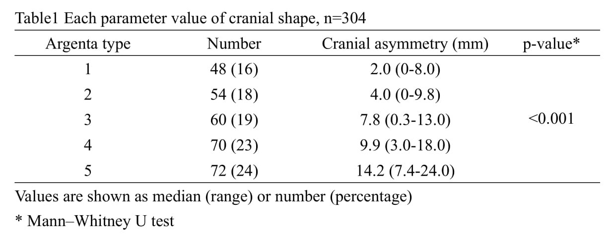
Design/Methods: We conducted a prospective cohort study after obtaining the IRB approval. Study subjects were comprised of 304 healthy infants at age 3-7 months (mean: 4 months) who visited two medical centers in Japan from April 2020 to November 2021. Argenta classification defines DP as follows: Type 1: posterior asymmetry; Type 2: anterior ear shift on the side of the occipital flattening; Type 3: frontal asymmetry; Type 4: facial asymmetry; and Type 5 or severe: temporal bossing or posterior vertical cranial growth. CA values are expressed as the difference between two diagonal diameters located 30° from the sagittal line of the skull – with severe: defined as a CA \geq\ 12 mm. The correlation between classification types was analyzed by regression analysis. After determining DP severity using the Argenta classification, the median CA value was calculated. The frequency of CA < 12 mm and of Argenta classification type 5 was then determined.
Results: A strong correlation between Argenta classification and CA was found (r=0.72, p < 0.001). Of the 72 infants with Argenta classification type 5, 11 (15.2%) had CA ≤12 mm.Conclusion(s): CA values are more accurate in identifying the severity of DP than the Argenta classification system. However, if 3D scanning is unavailable, using the Argenta classification system is still of value.
Each parameter value of cranial shape

