Neonatal Neurology: Clinical
Category: Abstract Submission
Neurology 7: Neonatal Neurology Term Imaging
348 - Differences between Early versus Late Magnetic Resonance Imaging in Infants with Neonatal Encephalopathy Following Therapeutic Hypothermia
Monday, April 25, 2022
3:30 PM - 6:00 PM US MT
Poster Number: 348
Publication Number: 348.443
Publication Number: 348.443
Aisling A. Garvey, INFANT Research Centre, Boston, MA, United States; Hoda Elshibiny, Birgham and Women's Hospital, Boston, MA, United States; Terrie E. Inder, Harvard Medical School, Boston, MA, United States; Mohamed El-Dib, Harvard Medical School, Boston, MA, United States

Aisling A. Garvey, MB BCh BAO MRCPI
Neonatal Neurology Fellow
Brigham and Women's Hospital
Boston, Massachusetts, United States
Presenting Author(s)
Background: MRI is the gold standard diagnostic test to define the nature of brain injury in infants with Neonatal Encephalopathy (NE), with conventional MRI being of predictive value for subsequent neurodevelopmental outcome. Early diffusion weighted MRI may assist in delineating injury in the first days of life. However, as both clinical features and imaging findings evolve considerably over the first week of life, early imaging may not reflect the true extent of brain injury.
Objective: The aim of this study is to assess the value of combining early MRI with late MRI following therapeutic hypothermia for NE.
Design/Methods: A retrospective cohort study was conducted on term born infants who received therapeutic hypothermia for NE at the Brigham and Women’s Hospital between 2016-2020. We identified infants who had two MRIs: one within the first week of life (early) and one beyond the first week of life (late). All MRIs were clinically reported by pediatric neuroradiologists. Reports were categorized into normal or abnormal which included diffusion restriction, evidence of parenchymal or germinal matrix hemorrhage, extra-axial hemorrhage, or other signal abnormalities.
Results: 105 infants with NE were included (47 mild, 52 moderate, 6 severe). Median gestation age was 39.3 weeks (IQR 37.5 – 40.2) and median birth weight was 3.18kg (IQR 2.74 – 3.58). First MRI scan was performed at a median age of 4 days (IQR 4-4) with repeat scan at a median age of 15 days (IQR 11-24.5). A summary of MRI findings is outlined in Table 1. Twenty-three infants (22%) had a normal early scan of which 4 had injury noted on repeat MRI. (Figure 1) Eighty-two infants (78%) had abnormal findings noted on the early MRI, of which 7 had evolution of injury and 16 had complete resolution of findings. (Figure 2)Conclusion(s): In infants who received therapeutic hypothermia for NE, 26% had significant changes noted between their early and late MRIs. Relying solely on early MRI may overestimate injury in a proportion of infants and miss injury in another proportion. Caregivers should be aware of this limitation as it may impact prognostication. Combining early and late MRI following hypothermia allows for better characterization of brain injury.
Table 1. Summary of early and late MRI findings..png)
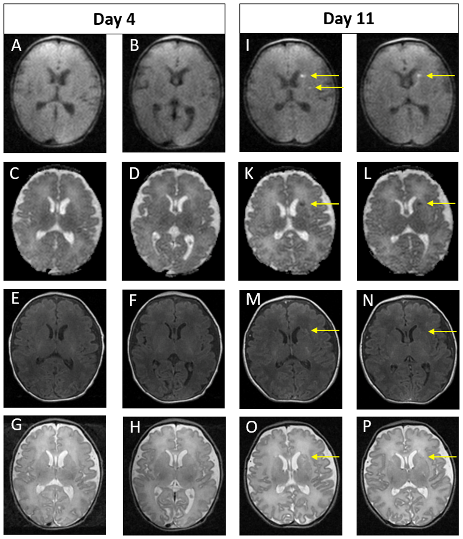
Figure 1. Example of MRI images of normal early scan and abnormal late scan. Images A-H on Day 4. Images I-P on Day 11.
In the late scan, punctate foci of diffusion restriction in the left caudate head and left putamen are seen on DWI (I and J) and on ADC (K and L). Punctate foci seen in left external capsule on axial T1 (M and N) and T2 (O and P) weighted images. These findings are not evident on the early scan (A – H).
Objective: The aim of this study is to assess the value of combining early MRI with late MRI following therapeutic hypothermia for NE.
Design/Methods: A retrospective cohort study was conducted on term born infants who received therapeutic hypothermia for NE at the Brigham and Women’s Hospital between 2016-2020. We identified infants who had two MRIs: one within the first week of life (early) and one beyond the first week of life (late). All MRIs were clinically reported by pediatric neuroradiologists. Reports were categorized into normal or abnormal which included diffusion restriction, evidence of parenchymal or germinal matrix hemorrhage, extra-axial hemorrhage, or other signal abnormalities.
Results: 105 infants with NE were included (47 mild, 52 moderate, 6 severe). Median gestation age was 39.3 weeks (IQR 37.5 – 40.2) and median birth weight was 3.18kg (IQR 2.74 – 3.58). First MRI scan was performed at a median age of 4 days (IQR 4-4) with repeat scan at a median age of 15 days (IQR 11-24.5). A summary of MRI findings is outlined in Table 1. Twenty-three infants (22%) had a normal early scan of which 4 had injury noted on repeat MRI. (Figure 1) Eighty-two infants (78%) had abnormal findings noted on the early MRI, of which 7 had evolution of injury and 16 had complete resolution of findings. (Figure 2)Conclusion(s): In infants who received therapeutic hypothermia for NE, 26% had significant changes noted between their early and late MRIs. Relying solely on early MRI may overestimate injury in a proportion of infants and miss injury in another proportion. Caregivers should be aware of this limitation as it may impact prognostication. Combining early and late MRI following hypothermia allows for better characterization of brain injury.
Table 1. Summary of early and late MRI findings.
.png)
Figure 1. Example of MRI images of normal early scan and abnormal late scan.
 Images A-H on Day 4. Images I-P on Day 11.
 In the late scan, punctate foci of diffusion restriction in the left caudate head and left putamen are seen on DWI (I and J) and on ADC (K and L). Punctate foci seen in left external capsule on axial T1 (M and N) and T2 (O and P) weighted images. These findings are not evident on the early scan (A – H).
