Neonatal Neurology: Clinical
Category: Abstract Submission
Neurology 4: Neonatal Neurology Preterm Clinical
465 - Elevated cerebral oxygen extraction is associated with impaired cerebral vasomotion in preterm infants
Sunday, April 24, 2022
3:30 PM - 6:00 PM US MT
Poster Number: 465
Zachary A. Vesoulis, Washington University School of Medicine, St. Louis, MO, United States; Halana V. Whitehead, Washington University in St. Louis School of Medicine, St. Louis, MO, United States; Devon Swofford, Washington University in St. Louis School of Medicine, Webster Groves, MO, United States; Steve M. Liao, Washington University in St. Louis School of Medicine, Saint Louis, MO, United States

Zachary A. Vesoulis, MD, MSCI
Assistant Professor
Washington University in St. Louis School of Medicine
St. Louis, Missouri, United States
Presenting Author(s)
Background: Vasomotion is the spontaneous, low-frequency, rhythmic contractions of arteries which play a role in facilitating tissue oxygen delivery. Computational analysis can identify the strength of these oscillations in systemic or cerebral blood flow. Although the preterm brain is at significant risk for injury from repeated hypoxia/ischemia, little is known about the relationship between vasomotion and cerebral oxygen extraction.
Objective: To identify the association between cerebral hypoxia/ischemia (defined as increasing cerebral oxygen extraction/decreasing cerebral saturation) and loss of cerebral or systemic vasomotion in preterm infants.
Design/Methods: Preterm infants born < 32 weeks with umbilical arterial lines were enrolled. Infants underwent continuous cerebral NIRS monitoring for the first 72h with simultaneous capture of mean arterial blood pressure (MABP) and pulse oximetry (SpO2). Simple error correction was performed to remove outliers and missing values.
Each recording was then divided into 10-min non-overlapping windows. The mean cerebral saturation (StO2) and fractional tissue oxygen extraction (FTOE, defined as SpO2-StO2/SpO2) was calculated for each window. Vasomotion strength of systemic blood flow (MABP) and a proxy for cerebral blood flow (StO2) were calculated using Welch’s power spectral density method in the frequency band 30-150 mHz for each 10-min window.
Vasomotion strength (spectral power) was then plotted against FTOE for systemic and cerebral vasomotion measures (Figure 2) for all 10-min windows with valid data. Vasomotion strength was also plotted against mean cerebral saturation (StO2, Figure 1). The strengths of these associations were then evaluated using standard linear fitting.
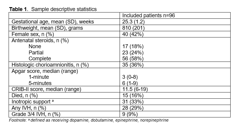
Results: 45187 ten-minute windows were captured from 96 infants were included with mean GA of 25.3 weeks, BW of 810g, 42% female, 29% had IVH, and 16% died during NICU hospitalization (Table 1).
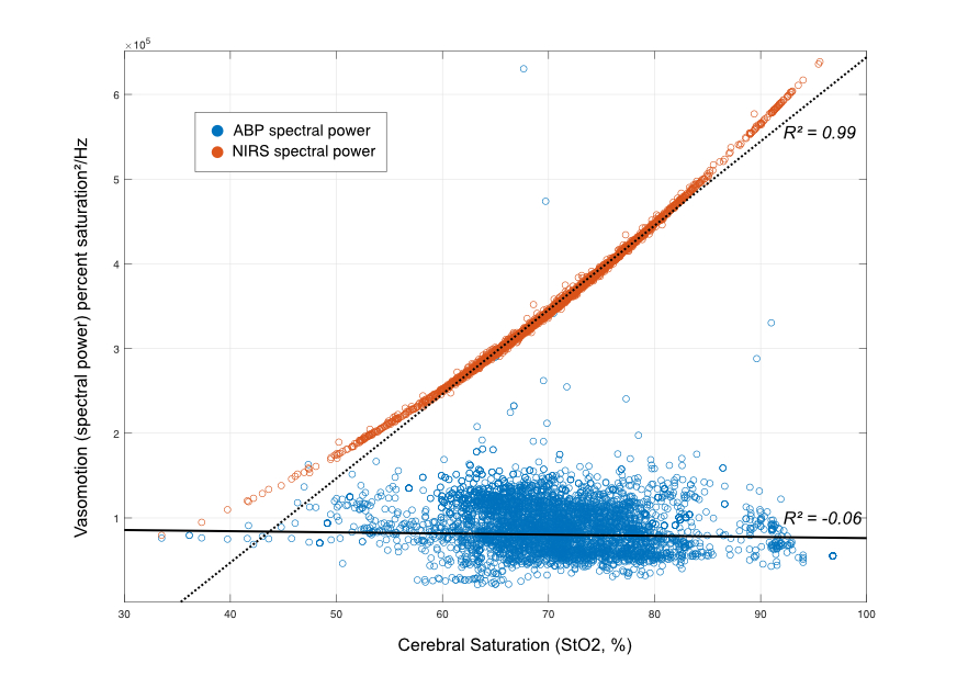
Systemic vasomotion (low-frequency oscillation in MABP) had no relationship to either cerebral oxygenation (R2= -0.06) or FTOE (R2=0.10). In contrast, a strong relationship was noted between cerebral vasomotion and cerebral oxygenation (R2=0.99) and cerebral FTOE (R2 -0.93) (Figures 1 and 2).Conclusion(s): As cerebral saturations dropped and FTOE increased (hypoxic-ischemia) there was a marked reduction in low-frequency cerebral vasomotion. The lack of a similar relationship with systemic vasomotion suggests that this is a localized phenomenon. Further research is needed to identify methods for recovering vasomotion to improve cerebral blood flow and decrease risk of hypoxia related brain injury.
Table 1 Sample descriptive statistics
Figure 1 The strength of vasomotion in the MABP (blue) and cerebral NIRS (orange) signals is shown on the y-axis and is plotted against the mean cerebral saturation for each valid data window on the x-axis. There is essentially no relationship between systemic vasomotion and cerebral saturation (solid line, R2 = -0.06). In comparison, there is a very strong relationship between increasing cerebral saturation and increasing cerebrovascular vasomotion (dashed line, R2 = 0.99).
Objective: To identify the association between cerebral hypoxia/ischemia (defined as increasing cerebral oxygen extraction/decreasing cerebral saturation) and loss of cerebral or systemic vasomotion in preterm infants.
Design/Methods: Preterm infants born < 32 weeks with umbilical arterial lines were enrolled. Infants underwent continuous cerebral NIRS monitoring for the first 72h with simultaneous capture of mean arterial blood pressure (MABP) and pulse oximetry (SpO2). Simple error correction was performed to remove outliers and missing values.
Each recording was then divided into 10-min non-overlapping windows. The mean cerebral saturation (StO2) and fractional tissue oxygen extraction (FTOE, defined as SpO2-StO2/SpO2) was calculated for each window. Vasomotion strength of systemic blood flow (MABP) and a proxy for cerebral blood flow (StO2) were calculated using Welch’s power spectral density method in the frequency band 30-150 mHz for each 10-min window.
Vasomotion strength (spectral power) was then plotted against FTOE for systemic and cerebral vasomotion measures (Figure 2) for all 10-min windows with valid data. Vasomotion strength was also plotted against mean cerebral saturation (StO2, Figure 1). The strengths of these associations were then evaluated using standard linear fitting.
Results: 45187 ten-minute windows were captured from 96 infants were included with mean GA of 25.3 weeks, BW of 810g, 42% female, 29% had IVH, and 16% died during NICU hospitalization (Table 1).
Systemic vasomotion (low-frequency oscillation in MABP) had no relationship to either cerebral oxygenation (R2= -0.06) or FTOE (R2=0.10). In contrast, a strong relationship was noted between cerebral vasomotion and cerebral oxygenation (R2=0.99) and cerebral FTOE (R2 -0.93) (Figures 1 and 2).Conclusion(s): As cerebral saturations dropped and FTOE increased (hypoxic-ischemia) there was a marked reduction in low-frequency cerebral vasomotion. The lack of a similar relationship with systemic vasomotion suggests that this is a localized phenomenon. Further research is needed to identify methods for recovering vasomotion to improve cerebral blood flow and decrease risk of hypoxia related brain injury.
Table 1
 Sample descriptive statistics
Figure 1
 The strength of vasomotion in the MABP (blue) and cerebral NIRS (orange) signals is shown on the y-axis and is plotted against the mean cerebral saturation for each valid data window on the x-axis. There is essentially no relationship between systemic vasomotion and cerebral saturation (solid line, R2 = -0.06). In comparison, there is a very strong relationship between increasing cerebral saturation and increasing cerebrovascular vasomotion (dashed line, R2 = 0.99).