Neonatal Neurology: Clinical
Category: Abstract Submission
Neurology 1: Clinical Pediatric & Neonatal Neurology
541 - Impaired global and regional brain growth in patients with isolated fetal ventriculomegaly
Friday, April 22, 2022
6:15 PM - 8:45 PM US MT
Poster Number: 541
Publication Number: 541.139
Publication Number: 541.139
Ha Tran, Children's National Health System, Washington, DC, United States; Nickie Andescavage, Children's National Health System, Washington, DC, United States; Todd C. Richmann, Children's National Health System, Washington, DC, United States; Kushal Kapse, Developing Brain Institute, Washington, DC, United States; Yushuf Sharker, Nationwide Children's Hospital, Washington, DC, United States; Jonathan Murnick, Children's National Health System, Washington, DC, United States; Gilbert Vezina, Children's National Health System, Washington, DC, United States; Adre du Plessis, George Washington University School of Medicine and Health Sciences, Washington, DC, United States; Catherine Limperopoulos, Children's National Health System, Washington, DC, United States

Ha Tran, MD
Pediatrics Resident
Children's National Health System
Washington, District of Columbia, United States
Presenting Author(s)
Background: Fetal ventriculomegaly (fVM) is one of the most common neurologic conditions identified in utero, with a wide variation in outcomes. Magnetic resonance imaging (MRI) has been shown to be superior to ultrasound to evaluate the fetal brain early in development, especially in the identification of additional brain injury and malformations that may adversely influence neurodevelopment. However, the prognosis for isolated fetal ventriculomegaly (ifVM) is equally challenging given the similarly wide variability in outcomes.
Objective: To compare quantitative global and regional brain volumes between fetuses with ifVM and healthy fetuses from uncomplicated pregnancies using volumetric quantitative MRI.
Design/Methods: This is a retrospective study of cases referred to the Prenatal Pediatrics Institute at Children’s National Health Center from November 2019 to December 2021 with ifVM at referral. Cases with additional brain or somatic malformations on fetal MRI were excluded. Using 3D reconstructions of T2-weighted MRI scans and semi-automated pipelines of fetal brain segmentations, we measured volumes for total brain volume (TBV), cerebrum, cerebellum, brainstem, along with cerebral cortical grey matter (CGM), white matter (WM) and subcortical grey matter (SCGM) and ventricular volumes. Descriptive statistics were used to characterize the cohorts with exploratory analyses in original and logarithmic scale and independent-sample t-tests.
Results: A total of 448 MRI (59 studies in ifVM, 389 reference controls) were collected, with gestation ages between 16-40 weeks (mean GA=30.5). As expected, the ifVM cohort had significantly enlarged lateral, 3rd and 4th ventricles compared to controls. Interestingly, cerebral volumes (mean ± SD of ifVM= 156cm3 ±84.4, controls= 168cm3 ± 84, p = 0.02), as well as cortical gray matter volumes (mean ± SD of ifVM= 62.9cm3 ±32.5, controls= 65.7cm3 ± 28.1, p < 0.01) were significantly reduced in ifVM compared to controls (Table 2).Conclusion(s): fVM is one of the most common anomalies of neurologic development detected on routine fetal screening with a wide range of developmental outcomes. Utilizing volumetric analysis with 3D MRI, we report in utero differences not only in ventricular volumes but in global and regional brain parenchymal volumes in fetuses with ifVM. The extent to which this cerebral growth impairment is associated with functional neurodevelopmental disabilities in fetuses with ifVM is unknown and currently under investigation.
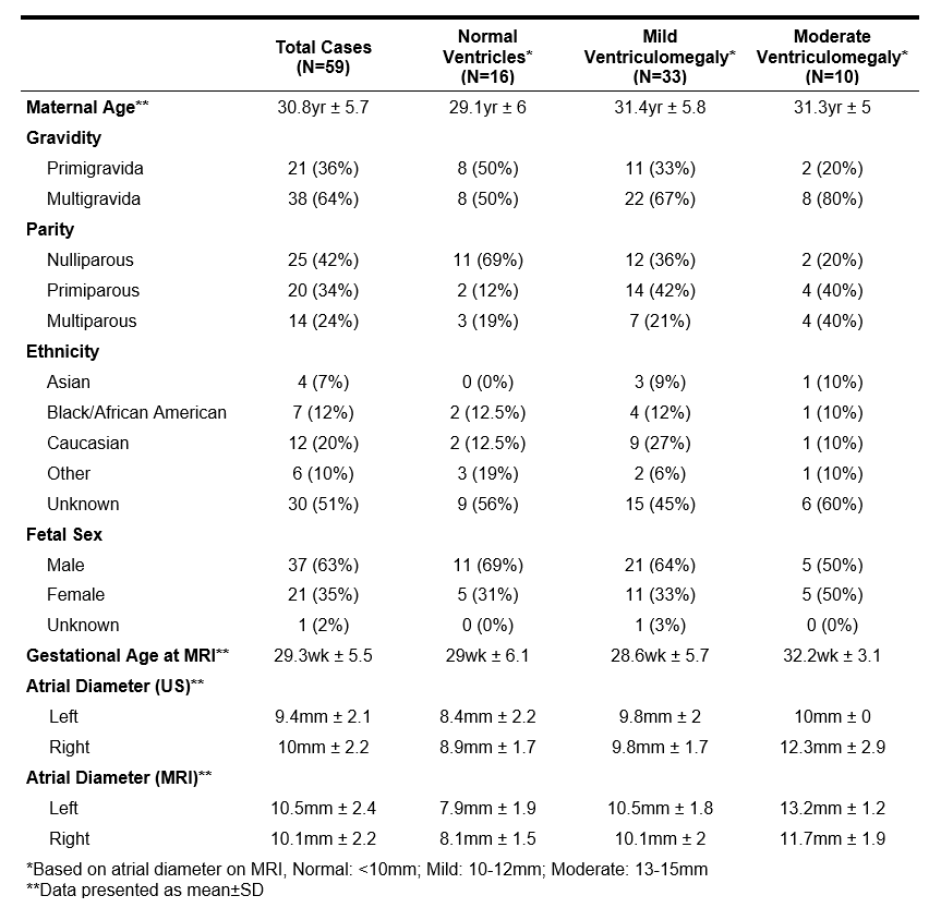
Table 1: Baseline characteristics of isolated fetal ventriculomegaly (ifVM) referrals
Table 2: Descriptive statistics for brain volumes (cm3).png)
Objective: To compare quantitative global and regional brain volumes between fetuses with ifVM and healthy fetuses from uncomplicated pregnancies using volumetric quantitative MRI.
Design/Methods: This is a retrospective study of cases referred to the Prenatal Pediatrics Institute at Children’s National Health Center from November 2019 to December 2021 with ifVM at referral. Cases with additional brain or somatic malformations on fetal MRI were excluded. Using 3D reconstructions of T2-weighted MRI scans and semi-automated pipelines of fetal brain segmentations, we measured volumes for total brain volume (TBV), cerebrum, cerebellum, brainstem, along with cerebral cortical grey matter (CGM), white matter (WM) and subcortical grey matter (SCGM) and ventricular volumes. Descriptive statistics were used to characterize the cohorts with exploratory analyses in original and logarithmic scale and independent-sample t-tests.
Results: A total of 448 MRI (59 studies in ifVM, 389 reference controls) were collected, with gestation ages between 16-40 weeks (mean GA=30.5). As expected, the ifVM cohort had significantly enlarged lateral, 3rd and 4th ventricles compared to controls. Interestingly, cerebral volumes (mean ± SD of ifVM= 156cm3 ±84.4, controls= 168cm3 ± 84, p = 0.02), as well as cortical gray matter volumes (mean ± SD of ifVM= 62.9cm3 ±32.5, controls= 65.7cm3 ± 28.1, p < 0.01) were significantly reduced in ifVM compared to controls (Table 2).Conclusion(s): fVM is one of the most common anomalies of neurologic development detected on routine fetal screening with a wide range of developmental outcomes. Utilizing volumetric analysis with 3D MRI, we report in utero differences not only in ventricular volumes but in global and regional brain parenchymal volumes in fetuses with ifVM. The extent to which this cerebral growth impairment is associated with functional neurodevelopmental disabilities in fetuses with ifVM is unknown and currently under investigation.
Table 1: Baseline characteristics of isolated fetal ventriculomegaly (ifVM) referrals

Table 2: Descriptive statistics for brain volumes (cm3)
.png)
