Neonatal Neurology: Clinical
Category: Abstract Submission
Neurology 6: Neonatal Neurology Preterm Imaging
341 - The Effect of Skin-To-Skin Care in Very Preterm Infants on Brain MRI Structural Abnormalities at Term-Equivalent Age
Monday, April 25, 2022
3:30 PM - 6:00 PM US MT
Poster Number: 341
Publication Number: 341.442
Publication Number: 341.442
Leah H. Fox, Cincinnati Children's Hospital Medical Center, CINCINNATI, OH, United States; Julia Kline, Cincinnati Children's Hospital Medical Center, Cincinnati, OH, United States; Chunyan Liu, Cincinnati Children's Hospital Medical Center, Cincinnati, OH, United States; Shelley Ehrlich, Cincinnati Children's Hospital Medical Center, Cincinnati, OH, United States; Nehal Parikh, Cincinnati Children's Hospital Medical Center, Cincinnati, OH, United States

Leah H. Fox, MD
Clinical Fellow
Cincinnati Children's Hospital Medical Center
CINCINNATI, Ohio, United States
Presenting Author(s)
Background: There is minimal research examining intermittent skin-to-skin contact (SSC) on neurodevelopmental outcomes in very preterm (VPT) infants. No studies have examined short-term effects on brain abnormalities on MRI.
Objective: Our objective was to determine the effects of duration of intermittent SSC on cortical surface area (primary) and additional measures of brain development (secondary) on term MRI in VPT infants.
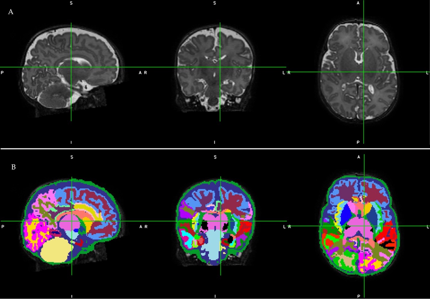
Design/Methods: This is a multicenter prospective cohort study of VPT infants born ≤32 weeks gestational age (GA). A 3T brain MRI was completed in the same scanner at 40-44 weeks postmenstrual age (PMA) and processed with the Developing Human Connectome Project (dHCP) pipeline to automatically calculate total cortical surface area, sulcal depth, gyrification index, curvature, and relative thalamic volume (Figure 1). We used linear mixed effects regression analyses to examine effects of SSC on dHCP metrics. To reduce confounding, propensity score model was used with CRIB II score, SSC during first week of life, antenatal steroids, PMA at time of MRI, intraventricular hemorrhage (IVH), indomethacin for prevention of IVH, and center. SSC was log-transformed to normalize the values.
Results: 246 infants had well-documented SSC. Mean (SD) GA was 29.3 (2.48) weeks and birth weight was 1342 (448) grams. 216 (88%) infants had ≥1 episode of SSC during NICU stay. The median (range) number of SSC episodes was 11 (3.25-23). 72% (n=117) experienced SCC in the first week, and median (range) time to first SSC was 2 (0-98) days. SSC exposure was significantly associated with maternal insurance, marital status, employment, and household income (p < 0.05) (Table 1). 210 infants had high-quality MRI images. SSC was not associated with cortical surface area (ß 1320.82; SE 1526.63; p=0.388). In secondary analysis, we observed a significant relationship between more SSC exposure and larger thalamic volume (ß 0.16; SE 0.07; p=0.0215) and increased curvature (ß 2.1; SE 0.97; p=0.032).Conclusion(s): In our cohort, SSC exposure was not associated with increased cortical maturation measures but was significantly associated with thalamic volume on MRI at term-equivalent age in VPT infants. This is the first study to correlate SSC and MRI metrics during infancy. SSC effects should be further correlated with cognitive, motor, and behavioral development at 2 years corrected age, as we are doing.
CVFox_CV.pdf
Figure 1 A representative participant’s raw T2-weighted midbrain MRI slices in sagittal, coronal, and axial orientations in panel A (top), and corresponding whole brain and regional cortical and tissue segmentations following automated processing using the Developing Human Connectome Project pipeline in panel B (bottom). Infant’s birth gestational age was 30.0 weeks and the MRI was performed at 41.1 weeks postmenstrual age. MRI Parameters: TE 166 ms, TR 18567 ms, FA 90°, voxel size 1.0×1.0× 1.0 mm3, and 3:43 minutes scan time.
Objective: Our objective was to determine the effects of duration of intermittent SSC on cortical surface area (primary) and additional measures of brain development (secondary) on term MRI in VPT infants.
Design/Methods: This is a multicenter prospective cohort study of VPT infants born ≤32 weeks gestational age (GA). A 3T brain MRI was completed in the same scanner at 40-44 weeks postmenstrual age (PMA) and processed with the Developing Human Connectome Project (dHCP) pipeline to automatically calculate total cortical surface area, sulcal depth, gyrification index, curvature, and relative thalamic volume (Figure 1). We used linear mixed effects regression analyses to examine effects of SSC on dHCP metrics. To reduce confounding, propensity score model was used with CRIB II score, SSC during first week of life, antenatal steroids, PMA at time of MRI, intraventricular hemorrhage (IVH), indomethacin for prevention of IVH, and center. SSC was log-transformed to normalize the values.
Results: 246 infants had well-documented SSC. Mean (SD) GA was 29.3 (2.48) weeks and birth weight was 1342 (448) grams. 216 (88%) infants had ≥1 episode of SSC during NICU stay. The median (range) number of SSC episodes was 11 (3.25-23). 72% (n=117) experienced SCC in the first week, and median (range) time to first SSC was 2 (0-98) days. SSC exposure was significantly associated with maternal insurance, marital status, employment, and household income (p < 0.05) (Table 1). 210 infants had high-quality MRI images. SSC was not associated with cortical surface area (ß 1320.82; SE 1526.63; p=0.388). In secondary analysis, we observed a significant relationship between more SSC exposure and larger thalamic volume (ß 0.16; SE 0.07; p=0.0215) and increased curvature (ß 2.1; SE 0.97; p=0.032).Conclusion(s): In our cohort, SSC exposure was not associated with increased cortical maturation measures but was significantly associated with thalamic volume on MRI at term-equivalent age in VPT infants. This is the first study to correlate SSC and MRI metrics during infancy. SSC effects should be further correlated with cognitive, motor, and behavioral development at 2 years corrected age, as we are doing.
CVFox_CV.pdf
Figure 1
 A representative participant’s raw T2-weighted midbrain MRI slices in sagittal, coronal, and axial orientations in panel A (top), and corresponding whole brain and regional cortical and tissue segmentations following automated processing using the Developing Human Connectome Project pipeline in panel B (bottom). Infant’s birth gestational age was 30.0 weeks and the MRI was performed at 41.1 weeks postmenstrual age. MRI Parameters: TE 166 ms, TR 18567 ms, FA 90°, voxel size 1.0×1.0× 1.0 mm3, and 3:43 minutes scan time.