Neonatal Neurology: Clinical
Category: Abstract Submission
Neurology 4: Neonatal Neurology Preterm Clinical
471 - Sleep-state-dependent and region-specific development of brain functional connectivity in preterm infants
Sunday, April 24, 2022
3:30 PM - 6:00 PM US MT
Poster Number: 471
Publication Number: 471.342
Publication Number: 471.342
Anna Shiraki, Nagoya University Graduate School of Medicine, Nagoya, Aichi, Japan; Hiroyuki Kidokoro, Nagoya University, Nagoya, Aichi, Japan; Hama Watanabe, The University of Tokyo, Bunkyo, Tokyo, Japan; Gentaro Taga, The University of Tokyo, Bunkyo-ku, Tokyo, Japan; Hajime Narita, Nagoya University Graduate School of Medicine, nagoya, Aichi, Japan; Takamasa Mitsumatsu, Nagoya University Graduate School of Medicine, nagoya, Aichi, Japan; Sumire Kumai, Nagoya University Graduate School of Medicine, Kasugai, Aichi, Japan; Ryosuke Suzui, Nagoya University Graduate School of Medicine, Nagoya, Aichi, Japan; Fumi Sawamura, Nagoya University Graduate School of Medicine, Nagoya, Aichi, Japan; Masahiro Kawaguchi, Nagoya University Graduate School of Medicine, Nagoya, Aichi, Japan; Takeshi Suzuki, Nagoya University Graduate School of medicine, Nagoya, Aichi, Japan; Hiroyuki Yamamoto, Nagoya University Graduate School of Medicine, Nagoya, Aichi, Japan; Tomohiko Nakata, Nagoya University Graduate School of Medicine, Nagoya, Aichi, Japan; Yoshiaki Sato, Nagoya University Hospital, Nagoya, Aichi, Japan; Masahiro Hayakawa, Nagoya University Hospital, Nagoya, Aichi, Japan; Jun Natsume, Nagoya University Graduate School of Medicine, Nagoya, Aichi, Japan

Anna Shiraki, MD (she/her/hers)
Visiting researcher
Nagoya University Graduate School of Medicine
Nagoya, Aichi, Japan
Presenting Author(s)
Background: The development of functional connectivity networks (FCNs) in preterm infants is not well understood.
Objective: Developmental changes in FCNs were assessed during sleep states in various brain regions in preterm infants using simultaneous electroencephalography (EEG) and functional near-infrared spectroscopy (NIRS).
Design/Methods: Between 2020 and 2021, we assessed 87 recordings from 37 preterm infants (gestational age ≤ 34 weeks) without severe brain injury. EEG was performed using a polygraph during active sleep (AS) and quiet sleep (QS), without sedation. An eight-channel NIRS device was placed around the infant’s head to detect slow changes ( < 0.1 Hz) in oxy- and deoxy-hemoglobin (Hb) concentrations. EEG, electrooculography, respiration pattern, and behavioral parameters were assessed every 30 s. The correlation coefficients of 28 channel pairs were calculated for the longest consecutive sequence in NIRS data during AS and QS and converted to z-scores using Fisher’s z-transformation to denote functional connectivity (FC) (Fig. 1). Linear regression analysis was used to assess associations among the average FC of all channels, FC associations between two channels, and differences in FC between AS and QS with postmenstrual age (PMA).
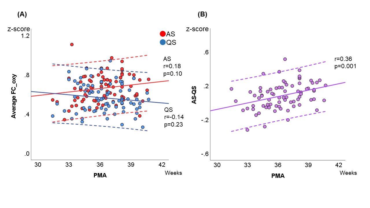
Results: The median (range) gestational age at birth and at EEG-functional NIRS recording were 32.5 (24.6–34.9) and 36.4 (31.4–40.7) weeks, respectively. Average FC based on oxy-Hb tended to increase at 40 weeks PMA during AS and to decrease during QS. The difference in average FC during AS and QS increased with PMA (P = 0.001) (Fig. 2). The trend in deoxy-Hb concentrations was similar. Three region-specific developmental FC patterns were observed in each pair of channels. FC between frontal and occipital areas and between inter-hemispheric temporal areas increased during AS and was stable during QS. This pattern reflected that of the average FC during AS and QS. FC between inter-hemispheric frontal areas was stable at high levels during AS and increased during QS. In these areas, FC increased with PMA during QS until it was similar to that during AS. FC between the inter-hemispheric occipital areas increased slightly during both AS and QS. In addition to the developmental FC pattern, FC strength in each channel pair differed among brain regions (Fig. 3).Conclusion(s): FCNs develop in preterm infants in a sleep-state-dependent and brain-region-related manner at 30–40 weeks PMA. Further studies are needed to determine whether these developmental dynamics in preterm infants are associated with later neurodevelopmental outcomes.
Biography20211231.pdf
Figure 2. Developmental changes in functional connectivity. Scatterplots showing (A) developmental changes in average functional connectivity (FC) of the 28 channel pairs during active sleep (AS, red) and quiet sleep (QS, blue) based on oxy-Hb concentrations, and (B) the average FC differences between AS and QS (purple) of the 28 channel pairs. The solid lines indicate the regression line, and the dotted lines represent the 95% confidence interval.
FC, functional connectivity; AS, active sleep; QS, quiet sleep; PMA, postmenstrual age.
Objective: Developmental changes in FCNs were assessed during sleep states in various brain regions in preterm infants using simultaneous electroencephalography (EEG) and functional near-infrared spectroscopy (NIRS).
Design/Methods: Between 2020 and 2021, we assessed 87 recordings from 37 preterm infants (gestational age ≤ 34 weeks) without severe brain injury. EEG was performed using a polygraph during active sleep (AS) and quiet sleep (QS), without sedation. An eight-channel NIRS device was placed around the infant’s head to detect slow changes ( < 0.1 Hz) in oxy- and deoxy-hemoglobin (Hb) concentrations. EEG, electrooculography, respiration pattern, and behavioral parameters were assessed every 30 s. The correlation coefficients of 28 channel pairs were calculated for the longest consecutive sequence in NIRS data during AS and QS and converted to z-scores using Fisher’s z-transformation to denote functional connectivity (FC) (Fig. 1). Linear regression analysis was used to assess associations among the average FC of all channels, FC associations between two channels, and differences in FC between AS and QS with postmenstrual age (PMA).
Results: The median (range) gestational age at birth and at EEG-functional NIRS recording were 32.5 (24.6–34.9) and 36.4 (31.4–40.7) weeks, respectively. Average FC based on oxy-Hb tended to increase at 40 weeks PMA during AS and to decrease during QS. The difference in average FC during AS and QS increased with PMA (P = 0.001) (Fig. 2). The trend in deoxy-Hb concentrations was similar. Three region-specific developmental FC patterns were observed in each pair of channels. FC between frontal and occipital areas and between inter-hemispheric temporal areas increased during AS and was stable during QS. This pattern reflected that of the average FC during AS and QS. FC between inter-hemispheric frontal areas was stable at high levels during AS and increased during QS. In these areas, FC increased with PMA during QS until it was similar to that during AS. FC between the inter-hemispheric occipital areas increased slightly during both AS and QS. In addition to the developmental FC pattern, FC strength in each channel pair differed among brain regions (Fig. 3).Conclusion(s): FCNs develop in preterm infants in a sleep-state-dependent and brain-region-related manner at 30–40 weeks PMA. Further studies are needed to determine whether these developmental dynamics in preterm infants are associated with later neurodevelopmental outcomes.
Biography20211231.pdf
Figure 2. Developmental changes in functional connectivity.
 Scatterplots showing (A) developmental changes in average functional connectivity (FC) of the 28 channel pairs during active sleep (AS, red) and quiet sleep (QS, blue) based on oxy-Hb concentrations, and (B) the average FC differences between AS and QS (purple) of the 28 channel pairs. The solid lines indicate the regression line, and the dotted lines represent the 95% confidence interval.
FC, functional connectivity; AS, active sleep; QS, quiet sleep; PMA, postmenstrual age.
