Neonatal General
Category: Abstract Submission
Neonatology General 4
388 - Cranial morphology reference values for based on 3D scan analysis in 1-month-old healthy Japanese infants
Friday, April 22, 2022
6:15 PM - 8:45 PM US MT
Poster Number: 388
Publication Number: 388.134
Publication Number: 388.134
Nobuhiko Nagano, Department of Pediatrics and Child Health, Nihon University School of Medicine, Tokyo, Tokyo, Japan; Hiroshi Miyabayashi, Nihon University school of medicine, Kasukabe, Saitama, Japan; Risa Kato, Department of Pediatrics and Child Health, Nihon University School of Medicine, Itabashi-ku, Tokyo, Japan; Takanori Noto, Nihon University Itabashi Hospital, nerima hikawadai 3-40-6, Tokyo, Japan; Shin Hashimoto, Kasukabe Medical Center, Kasukabe-shi, Saitama, Japan; Katsuya Saito, no, Kasukabe, Saitama, Japan; Ichiro Morioka, Nihon University School of Medicine, Itabashi, Tokyo, Japan

Hiroshi Miyabayashi, None
MD
Kasukabe medical center
Kasukabe, Saitama, Japan
Presenting Author(s)
Background: Three-dimensional (3D) scanners are widely used in the US to determine the cranial shape of infants and to guide treatment for deformational plagiocephaly (DP). Reference values of cranial morphology in Japanese infants and the prevalence of DP have not been established in Japan.
Objective: To establish reference values for cranial morphology in 1-month-old Japanese infants using 3D scanning and to determine the prevalence of DP.
Design/Methods: 1-month-old healthy Japanese infants who visited three hospitals from April 2020 to April 2021 were enrolled. Low birthweight or preterm infants and infants with neonatal asphyxia (5-min Apgar score < 7) were excluded. Cranial shape was measured using a 3D scanner (Artec Eva) and was analyzed using image analysis software. Cranial vault asymmetry index (CVAI) >3.5% or ≥10% were used as criteria for diagnosing DP or severe DP, respectively.
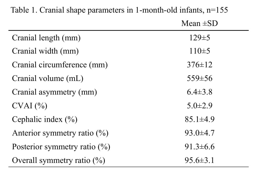
Results: A total of 155 participants (84 boys, 71 girls) was enrolled with mean gestational age (GA) of 39±1 wks and birthweight (BW) of 3,090±332 g. Mean age and weight at measurement were 35.7±6.3 days and 4,344 ± 500 g, respectively. Table 1 shows the mean values of each parameter. The prevalence of DP or severe DP was 65.8% or 5.8%, respectively. Sex differences were observed for cranial width, circumference, and volume.Conclusion(s): We were able to establish reference values for cranial morphology using 3D scan analysis in 1-month-old healthy Japanese infants. We found that the prevalence of DP and severe DP was 65% and 5%, respectively.
Cranial shape parameters in 1-month-old infants

