Neonatal Neurology: Basic
Category: Abstract Submission
Neurology 3: Basic-Translational
414 - Increased molecular interactions between IAIPs and HMGB1 in the neonatal brain after exposure to hypoxic-ischemic injury
Saturday, April 23, 2022
3:30 PM - 6:00 PM US MT
Poster Number: 414
Publication Number: 414.237
Publication Number: 414.237
Katie Gu, Women & Infants Hospital of Rhode Island, Rohnert Park, CA, United States; Saira Moazzam, Women & Infants Hospital of Rhode Island, Providence, RI, United States; Yow-Pin Lim, ProThera Biologics, Inc., Providence, RI, United States; Barbara Stonestreet, Women & Infants Hospital of Rhode Island, Providence, RI, United States; Xiaodi Chen, The Warren Alpert Medical School of Brown University, Providence, RI, United States

Katie Gu
n/a
Women & Infants Hospital of Rhode Island
Rohnert Park, California, United States
Presenting Author(s)
Background: The high mobility group box-1 (HMGB1) is a damage-associated molecular pattern protein that stimulates inflammatory cytokines and accentuates neuroinflammation after release from damaged cells. On the other hand, Inter-alpha inhibitor proteins (IAIPs) are naturally occurring immunomodulatory molecules that inhibit pro-inflammatory cytokines and exert neuroprotective effects on HI brain injury. We have shown that HMGB1 and IAIPs bind directly in vitro, and HMGB1 increases and co-localizes with IAIPs in neonatal brains after HI. However, the potential for IAIP-HMGB1 molecular interactions after exposure to HI remains to be determined.
Objective: To test whether IAIPs interact with HMGB1 in situ in neonatal cerebral cortices after exposure to HI injury.
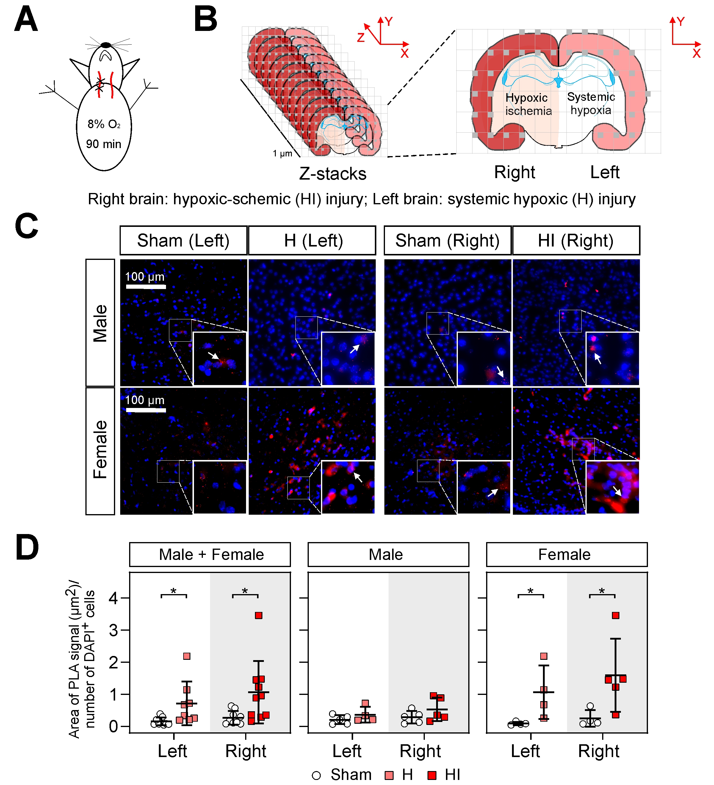
Design/Methods: Postnatal day 7 (P7) rats were assigned to groups: Sham and right carotid artery ligation and hypoxia (90 min of 8% O2, Fig. A). 72 h after HI, brains were perfused and fixed. On the left [hypoxia only (H) and Sham] and right (HI and Sham) parietal cortex, Proximity Ligation Assay® (PLA, Sigma-Aldrich) was used for detecting IAIPs-HMGB1 interactions. The parietal cortex was contoured, and a series of z-stack images were automatically acquired using a Systematic Random Sampling (SRS) method for offline, unbiased stereological analysis (Stereo Investigator, MBF Bioscience, Fig. B, PMID: 23348363). The z-stack images were combined as a maximum-intensity projection, and the quantification of PLA signal was plotted as the area of PLA signal per field (µm2)/number of DAPI+ cells (ImageJ/Fiji, NIH, PMID: 33044803). The data were log-transformed and compared between groups by Two-way ANOVA with the post-hoc Turkey HSD test.
Results: Representative PLA stains showed that PLA signals did not increase in left or right parietal cortices in males (Fig. C, upper panel), but increased in females (Fig. C, lower panel) after H and HI brain injury. Systemic H and HI resulted in significant increases in the IAIP-HMGB1 interactions in the parietal cortex in male+female and female rats (Fig. D, mean±SD, *P < 0.05). Statistical differences between H and HI were not observed.Conclusion(s): Systemic H and HI increased HI-related brain injury in neonatal rats in a sex-dependent manner. We speculate that increases in IAIP-HMGB1 interactions could facilitate some of the immunomodulatory and neuroprotective properties of IAIPs after HI brain injury in neonates.
PLA applied to cerebral cortices demonstrating increased IAIP-HMGB1 interactions after H and HI injury in situ. A) At P7, the right common carotid artery was ligated and followed by 90 min 8% of O2. Sham: male=5, female=5; HI: male= 4-5, female=4-5. B) At P10, a series of z-stack cortical images were acquired using the SRS method. C) Representative PLA stains showed increased PLA signals in female neonatal rats after exposure to H and HI injuries, but not in male neonatal rats, compared to the Sham. D) Results are represented as dot plots (mean±SD, *P < 0.05).
Objective: To test whether IAIPs interact with HMGB1 in situ in neonatal cerebral cortices after exposure to HI injury.
Design/Methods: Postnatal day 7 (P7) rats were assigned to groups: Sham and right carotid artery ligation and hypoxia (90 min of 8% O2, Fig. A). 72 h after HI, brains were perfused and fixed. On the left [hypoxia only (H) and Sham] and right (HI and Sham) parietal cortex, Proximity Ligation Assay® (PLA, Sigma-Aldrich) was used for detecting IAIPs-HMGB1 interactions. The parietal cortex was contoured, and a series of z-stack images were automatically acquired using a Systematic Random Sampling (SRS) method for offline, unbiased stereological analysis (Stereo Investigator, MBF Bioscience, Fig. B, PMID: 23348363). The z-stack images were combined as a maximum-intensity projection, and the quantification of PLA signal was plotted as the area of PLA signal per field (µm2)/number of DAPI+ cells (ImageJ/Fiji, NIH, PMID: 33044803). The data were log-transformed and compared between groups by Two-way ANOVA with the post-hoc Turkey HSD test.
Results: Representative PLA stains showed that PLA signals did not increase in left or right parietal cortices in males (Fig. C, upper panel), but increased in females (Fig. C, lower panel) after H and HI brain injury. Systemic H and HI resulted in significant increases in the IAIP-HMGB1 interactions in the parietal cortex in male+female and female rats (Fig. D, mean±SD, *P < 0.05). Statistical differences between H and HI were not observed.Conclusion(s): Systemic H and HI increased HI-related brain injury in neonatal rats in a sex-dependent manner. We speculate that increases in IAIP-HMGB1 interactions could facilitate some of the immunomodulatory and neuroprotective properties of IAIPs after HI brain injury in neonates.
PLA applied to cerebral cortices demonstrating increased IAIP-HMGB1 interactions after H and HI injury in situ.
 A) At P7, the right common carotid artery was ligated and followed by 90 min 8% of O2. Sham: male=5, female=5; HI: male= 4-5, female=4-5. B) At P10, a series of z-stack cortical images were acquired using the SRS method. C) Representative PLA stains showed increased PLA signals in female neonatal rats after exposure to H and HI injuries, but not in male neonatal rats, compared to the Sham. D) Results are represented as dot plots (mean±SD, *P < 0.05).