Neonatal Neurology: Basic
Category: Abstract Submission
Neurology 3: Basic-Translational
418 - Rectal temperature 1h after inflammation-sensitized hypoxia-ischemia predicts long-term white matter and cortical pathology in the late preterm ferret
Saturday, April 23, 2022
3:30 PM - 6:00 PM US MT
Poster Number: 418
Publication Number: 418.237
Publication Number: 418.237
Olivia R. White, University of Washington School of Medicine, Seattle, WA, United States; Kylie Corry, University of Washington School of Medicine, Seattle, WA, United States; Daniel Moralejo, University of Washington, Seattle, WA, United States; Janessa Law, University of Washington School of Medicine, Seattle, WA, United States; Jessica M. Snyder, University of Washington, Seattle, WA, United States; Ulrike Mietzsch, University of Washington School of Medicine, Seattle, WA, United States; Sandra E. Juul, University of Washington, Bellevue, WA, United States; Tommy Wood, University of Washington School of Medicine, Seattle, WA, United States

Thomas R. Wood, MD, PhD (he/him/his)
Assistant Professor
University of Washington School of Medicine
Seattle, Washington, United States
Presenting Author(s)
Background: Perinatal hypoxia-ischemia (HI) remains a common cause of neonatal mortality. The neurological corollaries of HI include a central injury pattern involving the basal ganglia and thalamus, which interferes with regulatory circuits involving temperature regulation. Spontaneous hypothermia occurs in both preclinical models and clinical cases of perinatal HI, and could act as a predictor for later neuropathological outcomes.
Objective: To determine whether spontaneous hypothermia in ferrets exposed to HI is associated with greater injury compared to normothermic littermates.
Design/Methods: Postnatal day (P) 17 ferrets were presensitized with 3 mg/kg of LPS before undergoing bilateral carotid artery (CA) ligation and a period of hypoxia-hyperoxia-hypoxia (30 min at 9%, 80%, 9%). The right CA ligation was reversed and animals were returned to the jills for 1h before rectal temperatures were gauged to establish post-HI nesting temperature (NT). A “normal” range for NT was determined as mean +/- 2 standard deviations NT in healthy control animals (34.4-37.4 °C, n=23). At P42, animals were euthanized and cortical development, ex vivo MRI, and neuropathology were quantitatively assessed. For statistical analyses, HI animals were separated into in-range (IR; >34.4°C) or below range (BR; < 34.4°C) groups.
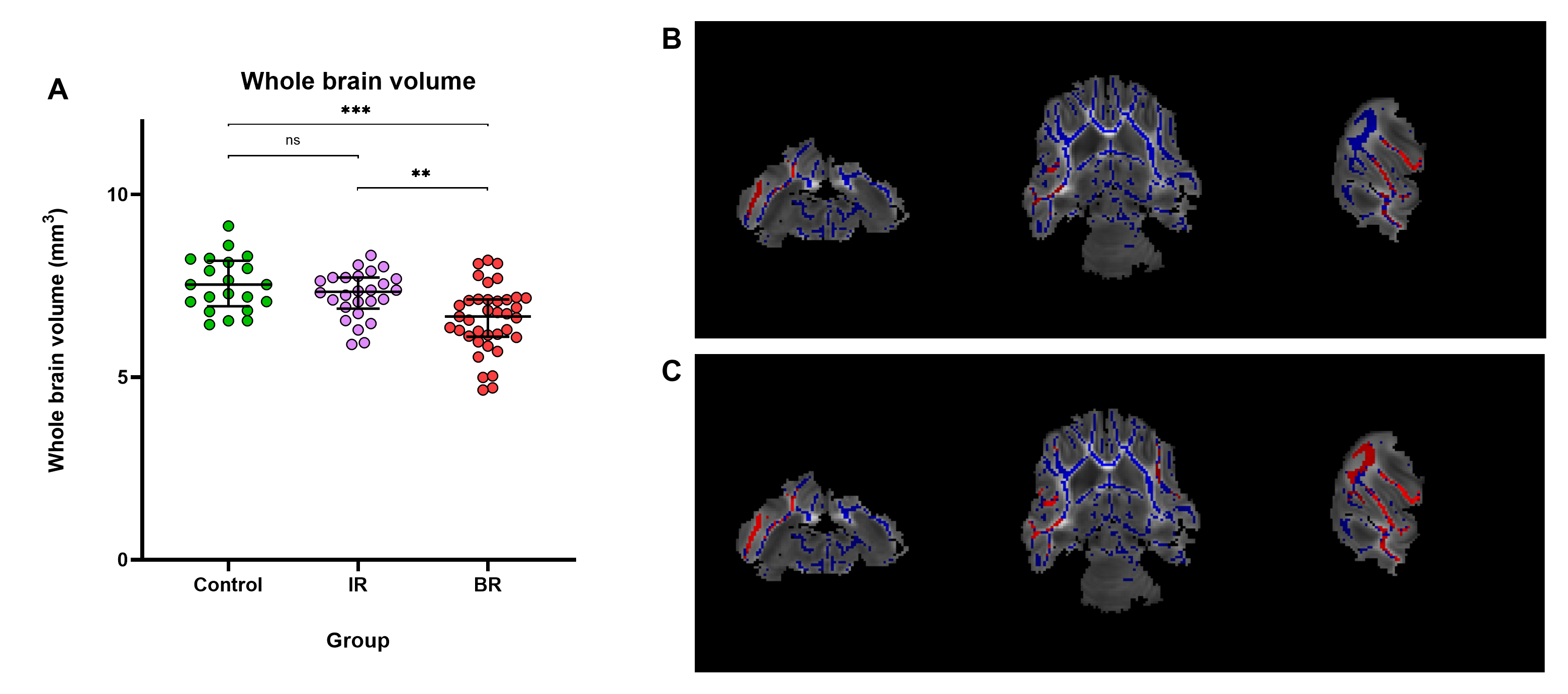
Results: Median (IQR) post-HI temperature of the IR (n=27) and BR (n=43) HI animals was 34.8°C (34.7-35.3°C) and 33.4°C (33.1-33.4°C), respectively. Whole brain volume in BR animals was significantly decreased compared to control and IR animals (Figure 1A).
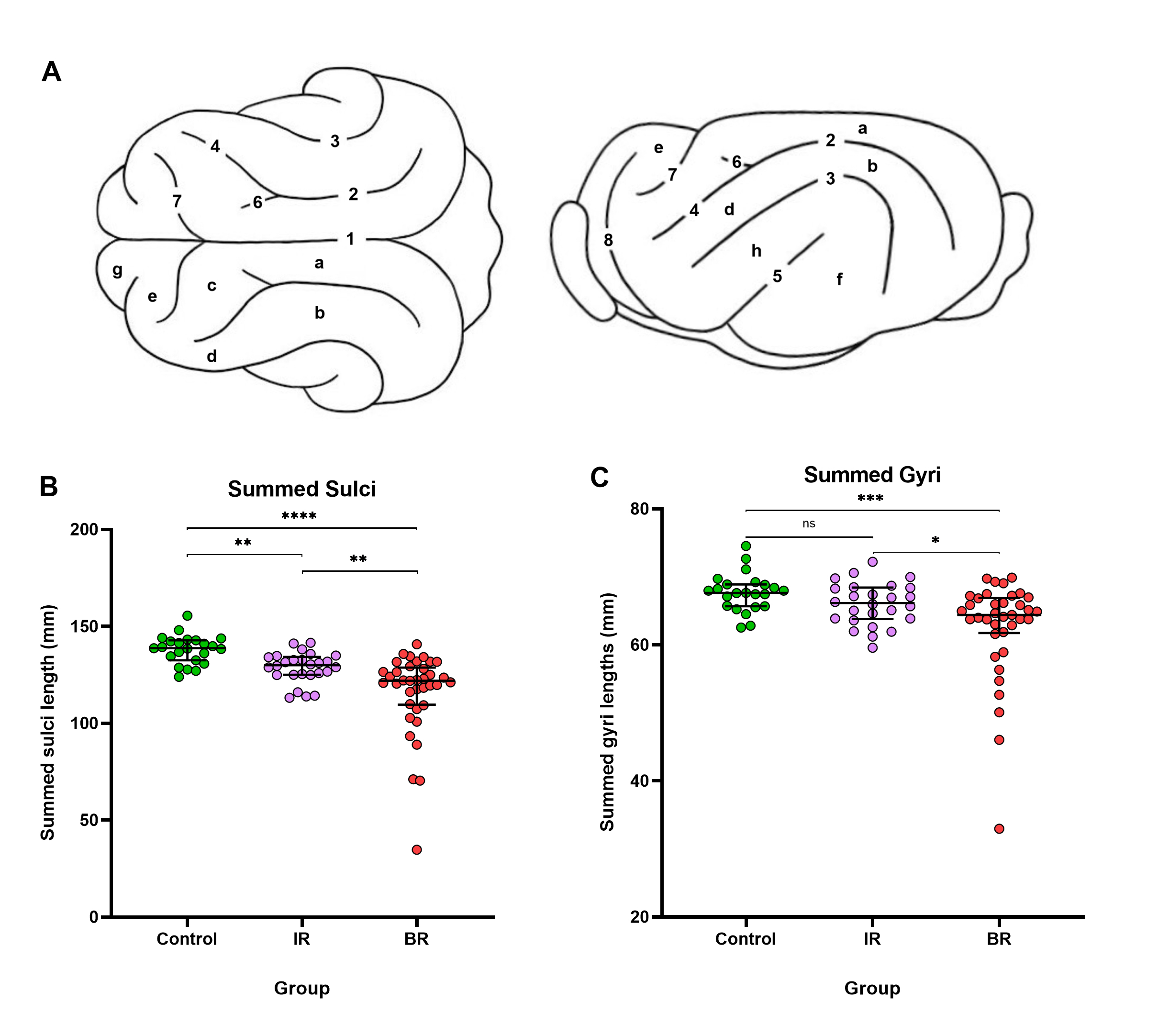
Fractional anisotropy was also reduced in BR brains compared to IR brains (Figure 1B/C). Morphological assessment showed BR brains had significantly narrower summed gyral measurements and shorter summed sulcal measurements than control and IR brains (Figure 2). Control and IR brains had lower cortical neuropathology scores compared to BR brains (Figure 3A). Quantitative immunohistochemistry showed increased GFAP staining of the thalamus and subcortical white matter in both IR and BR brains (Figure 3B/C), with BR brains displaying greater GFAP staining in the corpus callosum compared to IR brains (Figure 3D).Conclusion(s): Post-injury hypothermia in late preterm ferrets is associated with extensive cortical pathology and white matter changes, suggesting that post-HI NT can be utilized as an early biomarker to stratify injury severity and treatment in ferret studies of HI.
Figure 1. Magnetic Resonance and Diffusion Tensor Imaging. Comparisons of total brain volume and fractional anisotropy (FA) values throughout the white matter (blue). Red indicates significantly different FA values between IR and BR brains after threshold-free cluster enhancement (TFCE) adjustment for multiple comparisons. (A) The median and IQR are plotted on the graph. Analysis of whole brain volumetrics showed that BR brains had significantly decreased brain volumes when compared to IR and control brains. (B) At a significance level of 0.05, BR brains had significantly reduced FA unilaterally in the right posterior cerebral white matter tracts adjacent to the hippocampus. (C) At a significance level of 0.10, BR brains had significantly reduced FA bilaterally throughout the anterior and posterior cerebral white matter. ** Denotes p–value < 0.01, *** denotes p-value < 0.001, and ns denotes no significant difference.
Figure 2. Morphological Outcomes. The median and IQR are plotted on each graph. (A) Ferret brain atlas depicting: 1. Longitudinal Fissure, 2. Lateral Sulcus, 3. Suprasylvian Sulcus, 4. Coronal Sulcus, 5. Pseudosylvian Sulcus, 6. Ansinate Sulcus, 7. Cruciate Sulcus, 8. Presylvian Sulcus, a. Lateral Gyrus, b. Suprasylvian Gyrus, c. Posterior Sigmoid Gyrus, d. Coronal Gyrus, e. Ectosylvian Gyrus, g. Orbital Gyrus, i. Cerebellum exposed. (B) Summed sulci length in control brains compared to HI brains. After summing the length of each sulcus, IR and BR brains had significantly shorter sulci compared to control brains. IR brains displayed a significant improvement in summed totals of all sulcal lengths when compared to BR brains. (C) Summed gyri length in control brains relative to HI brains. BR brains had significantly narrower summed totals of all gyral widths when compared to IR and BR brains. * Denotes p–value < 0.05, ** denotes p–value < 0.01, *** denotes p-value < 0.001, **** denotes p-value < 0.0001, and ns denotes no significant difference.
Objective: To determine whether spontaneous hypothermia in ferrets exposed to HI is associated with greater injury compared to normothermic littermates.
Design/Methods: Postnatal day (P) 17 ferrets were presensitized with 3 mg/kg of LPS before undergoing bilateral carotid artery (CA) ligation and a period of hypoxia-hyperoxia-hypoxia (30 min at 9%, 80%, 9%). The right CA ligation was reversed and animals were returned to the jills for 1h before rectal temperatures were gauged to establish post-HI nesting temperature (NT). A “normal” range for NT was determined as mean +/- 2 standard deviations NT in healthy control animals (34.4-37.4 °C, n=23). At P42, animals were euthanized and cortical development, ex vivo MRI, and neuropathology were quantitatively assessed. For statistical analyses, HI animals were separated into in-range (IR; >34.4°C) or below range (BR; < 34.4°C) groups.
Results: Median (IQR) post-HI temperature of the IR (n=27) and BR (n=43) HI animals was 34.8°C (34.7-35.3°C) and 33.4°C (33.1-33.4°C), respectively. Whole brain volume in BR animals was significantly decreased compared to control and IR animals (Figure 1A).
Fractional anisotropy was also reduced in BR brains compared to IR brains (Figure 1B/C). Morphological assessment showed BR brains had significantly narrower summed gyral measurements and shorter summed sulcal measurements than control and IR brains (Figure 2). Control and IR brains had lower cortical neuropathology scores compared to BR brains (Figure 3A). Quantitative immunohistochemistry showed increased GFAP staining of the thalamus and subcortical white matter in both IR and BR brains (Figure 3B/C), with BR brains displaying greater GFAP staining in the corpus callosum compared to IR brains (Figure 3D).Conclusion(s): Post-injury hypothermia in late preterm ferrets is associated with extensive cortical pathology and white matter changes, suggesting that post-HI NT can be utilized as an early biomarker to stratify injury severity and treatment in ferret studies of HI.
Figure 1. Magnetic Resonance and Diffusion Tensor Imaging.
 Comparisons of total brain volume and fractional anisotropy (FA) values throughout the white matter (blue). Red indicates significantly different FA values between IR and BR brains after threshold-free cluster enhancement (TFCE) adjustment for multiple comparisons. (A) The median and IQR are plotted on the graph. Analysis of whole brain volumetrics showed that BR brains had significantly decreased brain volumes when compared to IR and control brains. (B) At a significance level of 0.05, BR brains had significantly reduced FA unilaterally in the right posterior cerebral white matter tracts adjacent to the hippocampus. (C) At a significance level of 0.10, BR brains had significantly reduced FA bilaterally throughout the anterior and posterior cerebral white matter. ** Denotes p–value < 0.01, *** denotes p-value < 0.001, and ns denotes no significant difference.
Figure 2. Morphological Outcomes.
 The median and IQR are plotted on each graph. (A) Ferret brain atlas depicting: 1. Longitudinal Fissure, 2. Lateral Sulcus, 3. Suprasylvian Sulcus, 4. Coronal Sulcus, 5. Pseudosylvian Sulcus, 6. Ansinate Sulcus, 7. Cruciate Sulcus, 8. Presylvian Sulcus, a. Lateral Gyrus, b. Suprasylvian Gyrus, c. Posterior Sigmoid Gyrus, d. Coronal Gyrus, e. Ectosylvian Gyrus, g. Orbital Gyrus, i. Cerebellum exposed. (B) Summed sulci length in control brains compared to HI brains. After summing the length of each sulcus, IR and BR brains had significantly shorter sulci compared to control brains. IR brains displayed a significant improvement in summed totals of all sulcal lengths when compared to BR brains. (C) Summed gyri length in control brains relative to HI brains. BR brains had significantly narrower summed totals of all gyral widths when compared to IR and BR brains. * Denotes p–value < 0.05, ** denotes p–value < 0.01, *** denotes p-value < 0.001, **** denotes p-value < 0.0001, and ns denotes no significant difference.