Breastfeeding/Human Milk
Category: Abstract Submission
Breastfeeding/Human Milk II
427 - Early breast milk exposure is associated with cortical maturation in preterm infants
Sunday, April 24, 2022
3:30 PM - 6:00 PM US MT
Poster Number: 427
Publication Number: 427.302
Publication Number: 427.302
Gemma Sullivan, University of Edinburgh, Edinburgh, Scotland, United Kingdom; Kadi Vaher, University of Edinburgh, Edinburgh, Scotland, United Kingdom; Manuel Blesa, University of Edinburgh, Edinburgh, Scotland, United Kingdom; Paola Galdi, MRC Centre for Reproductive Health, University of Edinburgh, UK, Edinburgh, Scotland, United Kingdom; David Q. Stoye, National Health Service, St. Albans, England, United Kingdom; Alan J. Quigley, NHS Lothian, Edinburgh, Scotland, United Kingdom; Michael Thrippleton, University of Edinburgh, Edinburgh, Scotland, United Kingdom; Mark Bastin, University of Edinburgh, Edinburgh, Scotland, United Kingdom; James P. Boardman, University of Edinburgh, Edinburgh, Scotland, United Kingdom
- GS
Gemma Sullivan, MBChB PhD
Dr
University of Edinburgh
Edinburgh, Scotland, United Kingdom
Presenting Author(s)
Background: Breast milk (BM) exposure is associated with improved neurocognitive outcomes following preterm birth but the neural substrates linking nutrition with outcome are uncertain. Neonatal MRI studies have shown that BM enhances white matter microstructure but effects on the developing cortex have not yet been described.
Objective: By combining nutritional data with brain MRI, we tested the hypothesis that high versus low BM exposure in preterm infants during neonatal care results in a cerebral cortical morphology that more closely resembles that of infants born at term.
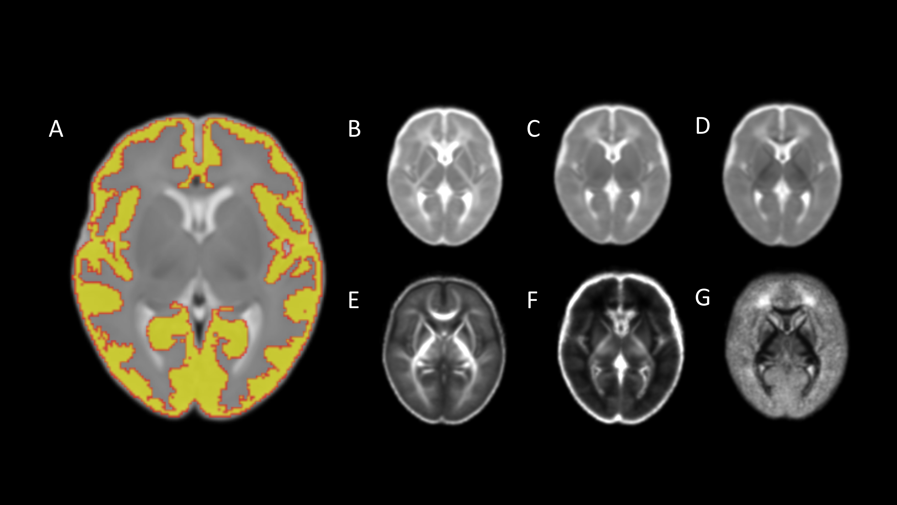
Design/Methods: We studied 135 preterm (mean GA 30+2 weeks, range 22+1 - 32+6) and 77 term-born infants (mean GA 39+4 weeks, range 36+3 - 42+1). Daily nutritional intake was collected from birth until hospital discharge to identify the proportion (%) of days preterm infants received exclusive BM feeds. Structural and diffusion MRI were performed at term-equivalent age. Cortical indices (volume, thickness, surface area, gyrification index, sulcal depth and curvature) and water diffusion parameters (fractional anisotropy (FA), mean diffusivity (MD), radial diffusivity (RD), axial diffusivity (AD), neurite density index (NDI), and orientation dispersion index (ODI)) were calculated. See figure 1 for representative brain maps. Imaging features residualised against age at MRI were compared between preterm infants who received exclusive BM for < 75% of inpatient days, preterm infants who received exclusive BM for ≥75% of inpatient days and healthy term-born controls.
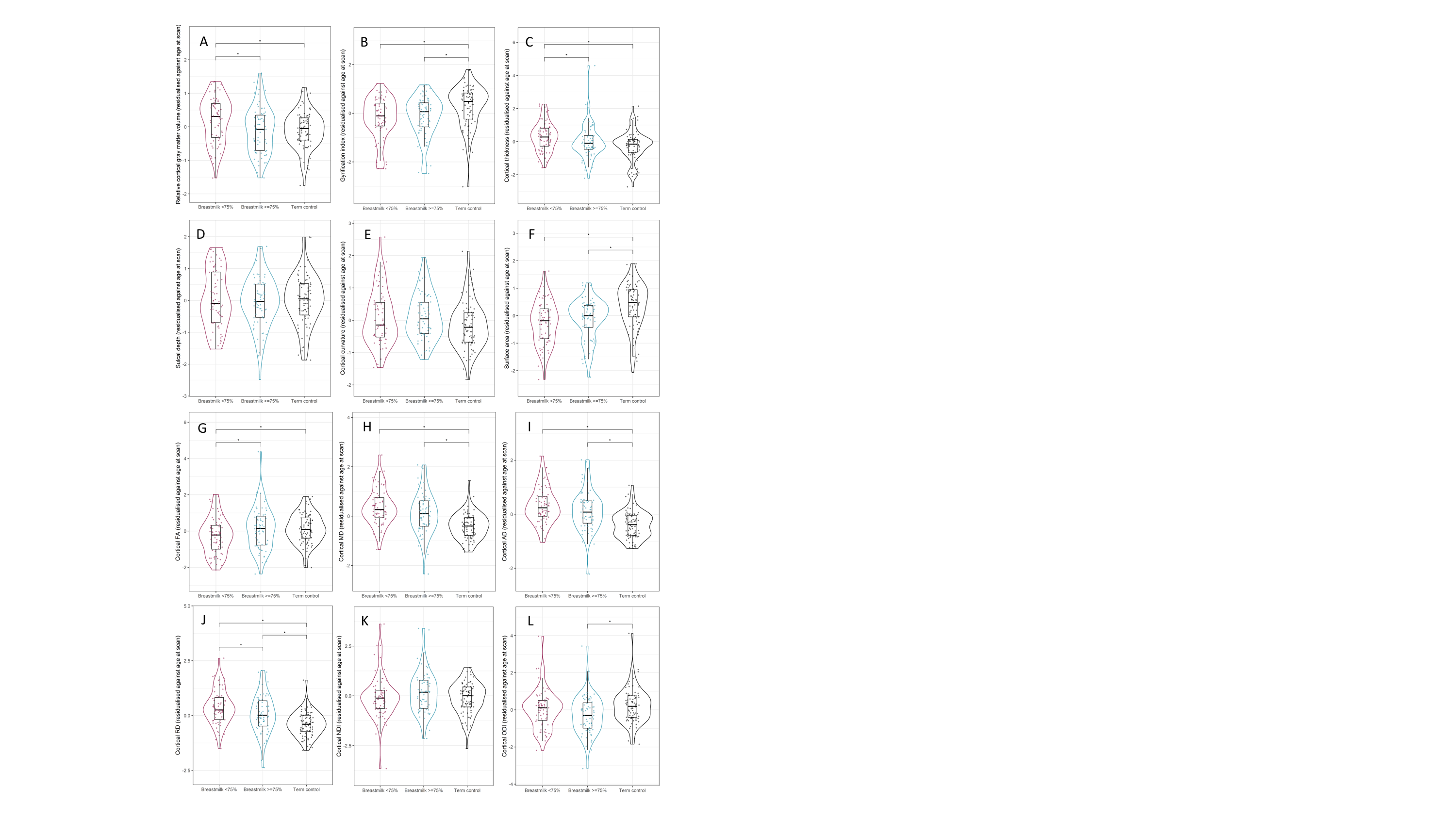
Results: 68 preterm infants received exclusive BM for < 75% of inpatient days and 67 preterm infants received exclusive BM for ≥75% of inpatient days. Participant characteristics are shown in Table 1. High compared with low BM exposure was associated with reduced cortical gray matter volume (d=0.47, p=0.014), thickness (d=0.42, p=0.039) and RD (d=0.38, p=0.039), and increased FA (d=0.38, p=0.037) after adjustment for age at MRI (Figure 2).The gyrification index, surface area, MD, AD and ODI were significantly different in preterm infants when compared to term-born controls but within the preterm group there was no effect of breast milk on these features.Conclusion(s): High versus low BM exposure in the weeks following preterm birth is associated with a cerebral cortical imaging phenotype that more closely resembles the brain morphology of healthy infants born at term. Breast milk may offer an intervention to optimise early cortical development following preterm birth, thereby reducing the risk of later neurocognitive impairment and psychiatric disease.
Figure 1. Representative brain maps. Averaged maps for (A) cortical segmentation, (B) axial diffusivity, (C) mean diffusivity, (D) radial diffusivity, (E) fractional anisotropy, (F) neurite density index, (G) orientation dispersion index.
Figure 2. Breast milk exposure and cortical maturation. Violin and box plots showing (A) relative cortical gray matter volume, (B) gyrification index, (C) cortical thickness, (D) sulcal depth, (E) cortical curvature, (F) surface area, (G) fractional anisotropy, (H) mean diffusivity, (I) axial diffusivity, (J) radial diffusivity, (K) neurite density index, (L) orientation dispersion index in preterm infants with low breast milk exposure (magenta), preterm infants with high breast milk exposure (blue) and term-born controls (black). *represents statistically significant pairwise comparisons after FDR correction.
Objective: By combining nutritional data with brain MRI, we tested the hypothesis that high versus low BM exposure in preterm infants during neonatal care results in a cerebral cortical morphology that more closely resembles that of infants born at term.
Design/Methods: We studied 135 preterm (mean GA 30+2 weeks, range 22+1 - 32+6) and 77 term-born infants (mean GA 39+4 weeks, range 36+3 - 42+1). Daily nutritional intake was collected from birth until hospital discharge to identify the proportion (%) of days preterm infants received exclusive BM feeds. Structural and diffusion MRI were performed at term-equivalent age. Cortical indices (volume, thickness, surface area, gyrification index, sulcal depth and curvature) and water diffusion parameters (fractional anisotropy (FA), mean diffusivity (MD), radial diffusivity (RD), axial diffusivity (AD), neurite density index (NDI), and orientation dispersion index (ODI)) were calculated. See figure 1 for representative brain maps. Imaging features residualised against age at MRI were compared between preterm infants who received exclusive BM for < 75% of inpatient days, preterm infants who received exclusive BM for ≥75% of inpatient days and healthy term-born controls.
Results: 68 preterm infants received exclusive BM for < 75% of inpatient days and 67 preterm infants received exclusive BM for ≥75% of inpatient days. Participant characteristics are shown in Table 1. High compared with low BM exposure was associated with reduced cortical gray matter volume (d=0.47, p=0.014), thickness (d=0.42, p=0.039) and RD (d=0.38, p=0.039), and increased FA (d=0.38, p=0.037) after adjustment for age at MRI (Figure 2).The gyrification index, surface area, MD, AD and ODI were significantly different in preterm infants when compared to term-born controls but within the preterm group there was no effect of breast milk on these features.Conclusion(s): High versus low BM exposure in the weeks following preterm birth is associated with a cerebral cortical imaging phenotype that more closely resembles the brain morphology of healthy infants born at term. Breast milk may offer an intervention to optimise early cortical development following preterm birth, thereby reducing the risk of later neurocognitive impairment and psychiatric disease.
Figure 1. Representative brain maps.
 Averaged maps for (A) cortical segmentation, (B) axial diffusivity, (C) mean diffusivity, (D) radial diffusivity, (E) fractional anisotropy, (F) neurite density index, (G) orientation dispersion index.
Figure 2. Breast milk exposure and cortical maturation.
 Violin and box plots showing (A) relative cortical gray matter volume, (B) gyrification index, (C) cortical thickness, (D) sulcal depth, (E) cortical curvature, (F) surface area, (G) fractional anisotropy, (H) mean diffusivity, (I) axial diffusivity, (J) radial diffusivity, (K) neurite density index, (L) orientation dispersion index in preterm infants with low breast milk exposure (magenta), preterm infants with high breast milk exposure (blue) and term-born controls (black). *represents statistically significant pairwise comparisons after FDR correction.