Neonatal Fetal Nutrition & Metabolism
Category: Abstract Submission
Neonatal Fetal Nutrition & Metabolism IV
457 - Using quantitative ultrasound to evaluate bone mineral density at birth and over time in infants born <30 weeks gestation
Sunday, April 24, 2022
3:30 PM - 6:00 PM US MT
Poster Number: 457
Publication Number: 457.335
Publication Number: 457.335
George Ziegler, Nationwide Children's Hospital, Columbus, OH, United States; Kenneth Jackson, The Ohio State University, Columbus, OH, United States; Laura Marzec, Nationwide Children's Hospital, Columbus, OH, United States; Rox Ann Sullivan, Nationwide Children's Hospital, Reynoldsburg, OH, United States; Erinn M. Hade, New York University Grossman School of Medicine, New York, NY, United States; Jonathan L. Slaughter, Nationwide Children's Hospital/The Ohio State University, Columbus, OH, United States

George Ziegler, MD
Neonatal-Perinatal Medicine Fellow
Nationwide Children's Hospital
Columbus, Ohio, United States
Presenting Author(s)
Background: Metabolic bone disease (MBD) remains an ongoing burden of prematurity and manifests with poor weight gain and growth failure, as well as fractures during infancy and childhood. Previous work has identified a relationship between birth weight and weight at 12 months of age and bone size and strength in the 6th and 7th decades of life.
Objective: To describe birth bone density in at-risk preterm infants and estimate the change in bone density from preterm birth to term or near-term postmenstrual age (PMA).
Design/Methods: Infants born 23-30 weeks gestation were enrolled in a prospective cohort study. To measure bone density, an initial quantitative bone ultrasound was collected within 7 days of birth using the BeamMed Sunlight Omnisense 9000; a follow up measurement was collected at 36-40 weeks PMA. Descriptive statistics such as frequencies, percentages, means, and standard deviations summarize infant characteristics and bone density measures; Pearson’s correlation coefficient estimates the relationship between the gestational age at birth measurement and the rate of change in bone density from birth to term or near-term PMA.
Results: 117 infants born 23-30 weeks gestation were enrolled. 106 infants completed an initial bone density measurement shortly after birth, and 77 infants completed a second measurement at 36-40 weeks PMA. A moderate positive correlation between birth gestational age (r=0.56) and birth weight (r=0.50) with initial bone density was identified. Compared to mean values of bone density identified by the manufacturer, our cohort exhibited lower bone density in more extremely preterm infants. From birth to term or near-term PMA, bone density decreased by an average of 1.5 m/s/day (95% CI: -2.1, -0.8), which did not vary substantially by sex (female: -1.6 m/s/day [95% CI: -2.4, -0.8], male: -1.2 m/s/day [95% CI: -2.2, -0.2]) or gestational age at birth (23-25 6/7wk: -1.7 m/s/day [95% CI: -3.1, -0.3], 26-27 6/7wk: -0.8 m/s/day [95% CI: -2.0, 0.4], 28-30wk: -1.8 m/s/day [95% CI: -2.8, -0.9]).Conclusion(s): The birth bone density of preterm infants increases with gestational age as well as with progressively greater birth weight. Infants born < 30 weeks gestation show a decrease in bone density over time as they progress to term or near-term PMA. We did not observe a clinically different pattern of longitudinal change in bone density relative to birth gestational age, differing from prior studies which identify a greater decrease in bone density among more extremely preterm infants.
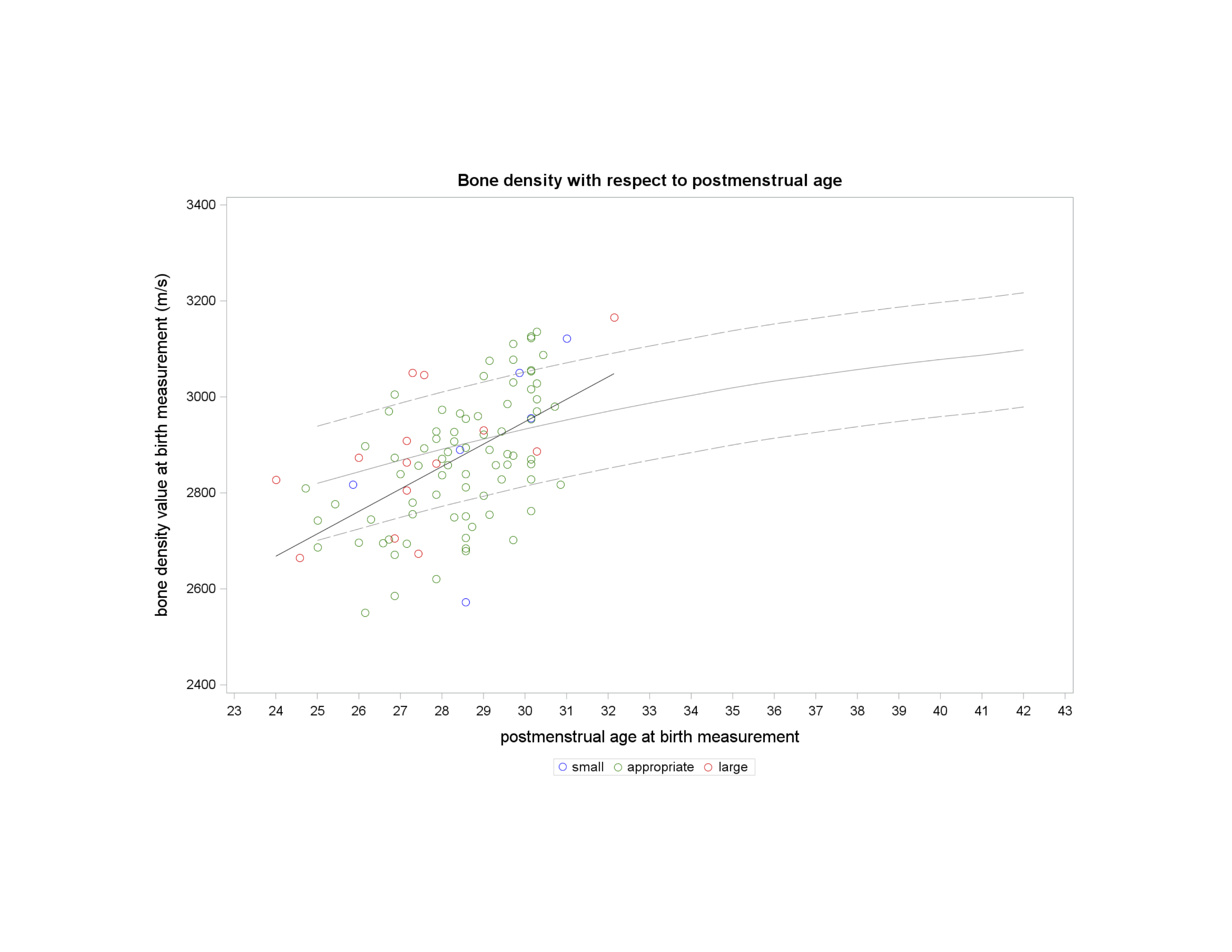
Figure 1. Bone density with respect to postmenstrual age Birth bone density displayed with respect to postmenstrual age at time of measurement. Note that the solid gray and dashed gray line represent mean bone density value and +/- one standard deviation provided by quantitative ultrasound manufacturer. Solid black line indicates mean of this study’s birth bone density value.
Small (Small for Gestational Age), Appropriate (Appropriate for Gestational Age), Large (Large for Gestational Age)
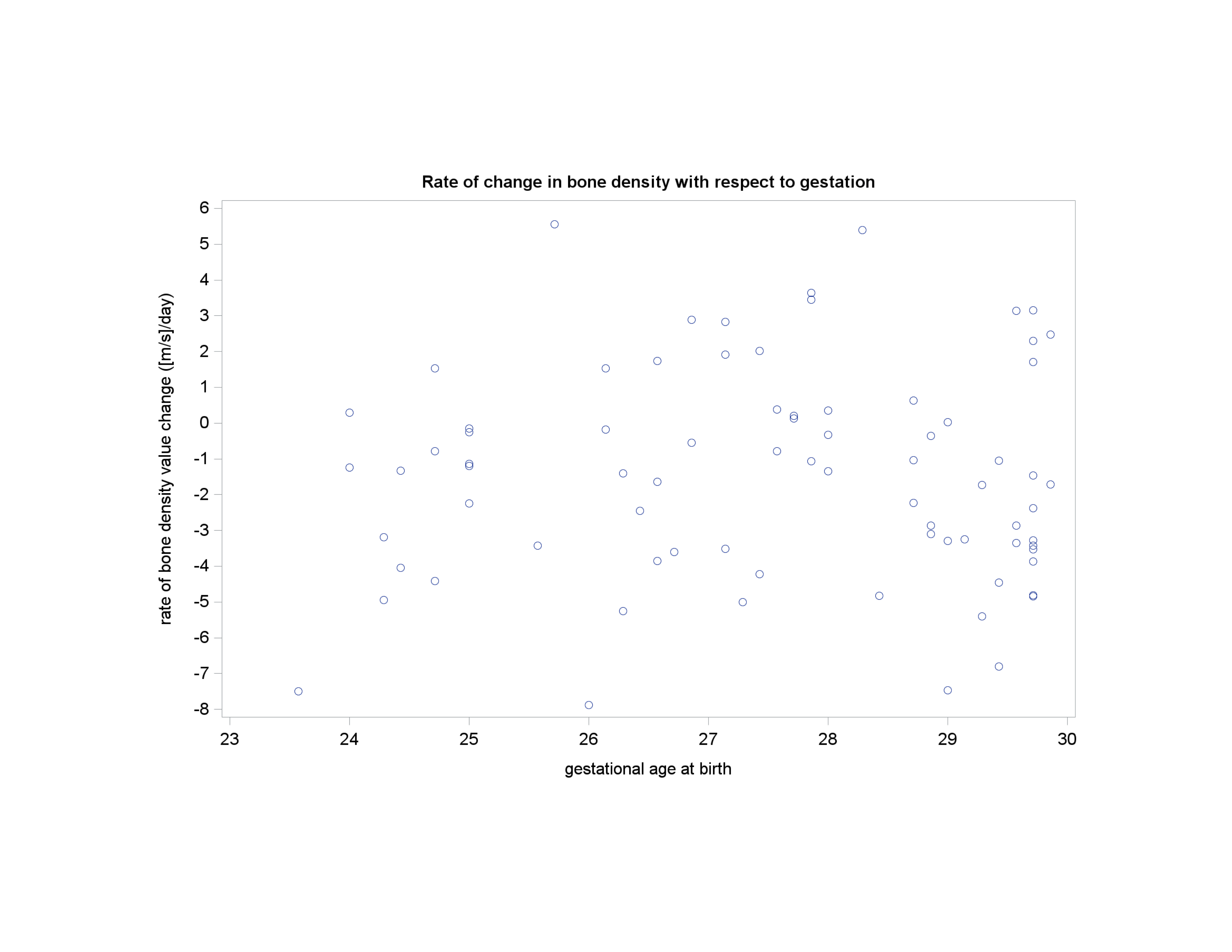
Figure 2. Rate of change in bone density with respect to gestation Rate of daily change in bone mineral density of individual study enrollees displayed with respect to gestational age at birth.
Objective: To describe birth bone density in at-risk preterm infants and estimate the change in bone density from preterm birth to term or near-term postmenstrual age (PMA).
Design/Methods: Infants born 23-30 weeks gestation were enrolled in a prospective cohort study. To measure bone density, an initial quantitative bone ultrasound was collected within 7 days of birth using the BeamMed Sunlight Omnisense 9000; a follow up measurement was collected at 36-40 weeks PMA. Descriptive statistics such as frequencies, percentages, means, and standard deviations summarize infant characteristics and bone density measures; Pearson’s correlation coefficient estimates the relationship between the gestational age at birth measurement and the rate of change in bone density from birth to term or near-term PMA.
Results: 117 infants born 23-30 weeks gestation were enrolled. 106 infants completed an initial bone density measurement shortly after birth, and 77 infants completed a second measurement at 36-40 weeks PMA. A moderate positive correlation between birth gestational age (r=0.56) and birth weight (r=0.50) with initial bone density was identified. Compared to mean values of bone density identified by the manufacturer, our cohort exhibited lower bone density in more extremely preterm infants. From birth to term or near-term PMA, bone density decreased by an average of 1.5 m/s/day (95% CI: -2.1, -0.8), which did not vary substantially by sex (female: -1.6 m/s/day [95% CI: -2.4, -0.8], male: -1.2 m/s/day [95% CI: -2.2, -0.2]) or gestational age at birth (23-25 6/7wk: -1.7 m/s/day [95% CI: -3.1, -0.3], 26-27 6/7wk: -0.8 m/s/day [95% CI: -2.0, 0.4], 28-30wk: -1.8 m/s/day [95% CI: -2.8, -0.9]).Conclusion(s): The birth bone density of preterm infants increases with gestational age as well as with progressively greater birth weight. Infants born < 30 weeks gestation show a decrease in bone density over time as they progress to term or near-term PMA. We did not observe a clinically different pattern of longitudinal change in bone density relative to birth gestational age, differing from prior studies which identify a greater decrease in bone density among more extremely preterm infants.
Figure 1. Bone density with respect to postmenstrual age
 Birth bone density displayed with respect to postmenstrual age at time of measurement. Note that the solid gray and dashed gray line represent mean bone density value and +/- one standard deviation provided by quantitative ultrasound manufacturer. Solid black line indicates mean of this study’s birth bone density value.
 Small (Small for Gestational Age), Appropriate (Appropriate for Gestational Age), Large (Large for Gestational Age)
Figure 2. Rate of change in bone density with respect to gestation
 Rate of daily change in bone mineral density of individual study enrollees displayed with respect to gestational age at birth.