Neonatal Pulmonology
Category: Abstract Submission
Neonatal Pulmonology I: Antenatal Exposures
424 - Ethanol exposure in a rodent model of preterm neonates leads to respiratory control dysfunction in female but not male rat pups
Monday, April 25, 2022
3:30 PM - 6:00 PM US MT
Poster Number: 424
Publication Number: 424.429
Publication Number: 424.429
Nicholas C. Rickman, Case Western Reserve University, Shaker Heights, OH, United States; Catherine Mayer, Case Western Reserve University School of Medicine, Cleveland, OH, United States; Sergei Vatolin, Case Western Reserve University School of Medicine, Cleveland, OH, United States; Cansu Tokat, Case Western Reserve University School of Medicine, Shaker Heights, OH, United States; Peter MacFarlane, Rainbow Babies & Children’s Hospital/ Case Western Reserve University, Cleveland, OH, United States; Cynthia F. Bearer, Case Western Reserve University School of Medicine, Cleveland, OH, United States
- NR
Nicholas C. Rickman, Bachelor's (he/him/his)
Research Assistant
Case Western Reserve University School of Medicine
Shaker Heights, Ohio, United States
Presenting Author(s)
Background: Fetal alcohol spectrum disorder (FASD) has been linked to numerous neurodevelopmental diseases, such as general hypoplasia, microcephaly, and several learning disorders.
Objective: Using a rodent model of preterm neonates, we investigated whether ethanol exposure disrupts central and peripheral mechanisms of respiratory neural control in neonatal rats, by assessing their ventilatory response to hypoxia and hypercapnia challenges.
Design/Methods: P5 female and male Sprague-Dawley rat pups received 0.8 or 4.4 mg/g of ethanol (or saline) delivered via IP injection. The ventilatory response to acute hypoxia (HVR; 10% O2, 5 min) and hypercapnia (HCVR; 5% CO2, 5 min) was measured using whole-body plethysmography 24 h later. Minute-by-minute changes in ventilation during hypoxic and hypercapnic challenge (expressed relative to baseline normoxic ventilation) were used as indices of respiratory control for the HVR and HCVR. Comparisons were made with saline injected rats.
Results: Female saline injected rats exhibited a pronounced biphasic hypoxic ventilatory response, increasing minute ventilation during the 1st and 2nd minute of hypoxia (0.71 ± 0.08 and 0.55 ± 0.05 ml/g/min, respectively). The lowest dose of ethanol (0.8 mg/g) had no effect on the biphasic (1st-2nd minute) HVR (Fig. 1), whereas it was abolished by 4.4 mg/g (1st-2nd min: 0.08 ± 0.11 – 0.05 ± 0.05 ml/g/min). The HCVR was also abolished by 4.4 mg/g of ethanol. Some mortality occurred in females (but not males) several days after (P7-P13) ethanol administration. Males appeared relatively unaffected by ethanol (Fig. 2).Conclusion(s): The effect on the early phase of the HVR implies functional impairment of peripheral mechanisms of neural control such as the carotid body chemoreceptors, while the attenuated hypercapnic response suggests deficits in central mechanisms. Such respiratory impairments and the associated mortality in females days after treatment could have implications for the increased risk of SIDS following ethanol exposure. The higher vulnerability of females compared to males may be related to sex-dependent differences in their ability to metabolize ethanol. Future studies will further qualify the degree of impact to respiratory control by ethanol, and elucidate the mechanism by which this impact occurs.
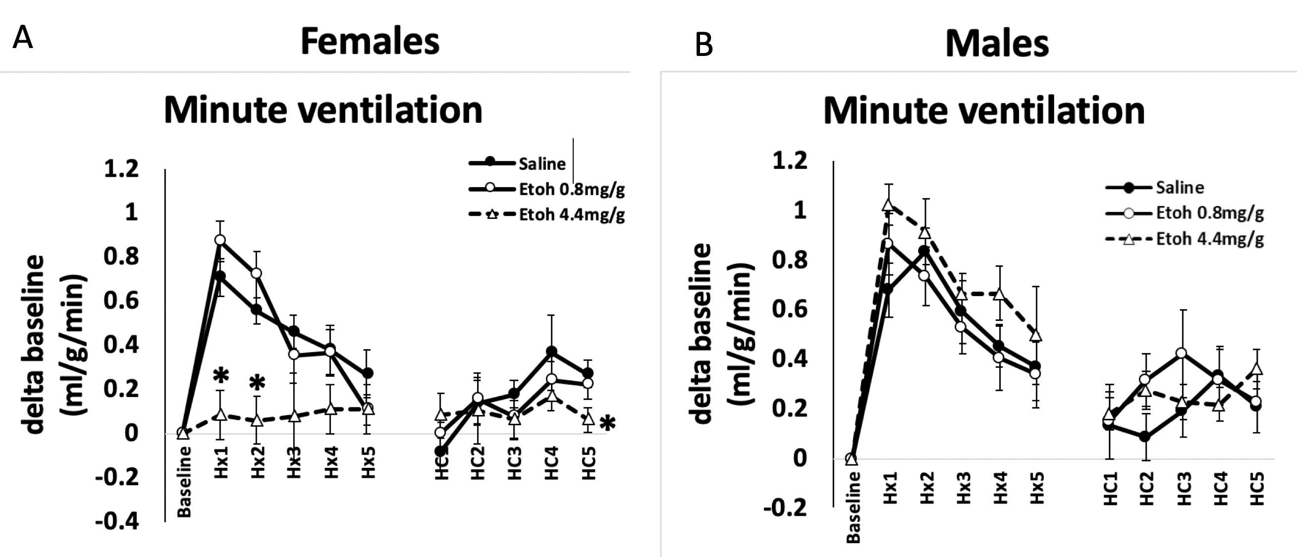
Hypoxic and Hypercapnic Ventilatory Response of P6 rat pups Ventilatory responses to acute hypoxia (HVR) and hypercapnia (HCVR) in 6 day old female (A) and male (B) rats 24 hours following an i.p. injection of ethanol (0.8mg/g or 4.4 mg/g) or saline. Note, both female and male rats exhibit an increase in minute ventilation during the first 1-2 minutes of hypoxia (Hx1-Hx2) followed by a progressive decline toward baseline (i.e. a biphasic HVR). In females (A), the highest dose of ethanol (4.4 mg/g) abolished the biphasic HVR and the 5th minute of the HCVR compared to saline treated rats. Males were not affected by ethanol. Values are expressed as means ± 1 S.E.M; the magnitude of the HVR and HCVR are expressed as a change (delta) in minute ventilation (ml/g/min) from baseline. *indicates significant difference from saline treated rats for a given time point in hypoxia (Hx) or hypercapnia (HC); P < 0.05.
Objective: Using a rodent model of preterm neonates, we investigated whether ethanol exposure disrupts central and peripheral mechanisms of respiratory neural control in neonatal rats, by assessing their ventilatory response to hypoxia and hypercapnia challenges.
Design/Methods: P5 female and male Sprague-Dawley rat pups received 0.8 or 4.4 mg/g of ethanol (or saline) delivered via IP injection. The ventilatory response to acute hypoxia (HVR; 10% O2, 5 min) and hypercapnia (HCVR; 5% CO2, 5 min) was measured using whole-body plethysmography 24 h later. Minute-by-minute changes in ventilation during hypoxic and hypercapnic challenge (expressed relative to baseline normoxic ventilation) were used as indices of respiratory control for the HVR and HCVR. Comparisons were made with saline injected rats.
Results: Female saline injected rats exhibited a pronounced biphasic hypoxic ventilatory response, increasing minute ventilation during the 1st and 2nd minute of hypoxia (0.71 ± 0.08 and 0.55 ± 0.05 ml/g/min, respectively). The lowest dose of ethanol (0.8 mg/g) had no effect on the biphasic (1st-2nd minute) HVR (Fig. 1), whereas it was abolished by 4.4 mg/g (1st-2nd min: 0.08 ± 0.11 – 0.05 ± 0.05 ml/g/min). The HCVR was also abolished by 4.4 mg/g of ethanol. Some mortality occurred in females (but not males) several days after (P7-P13) ethanol administration. Males appeared relatively unaffected by ethanol (Fig. 2).Conclusion(s): The effect on the early phase of the HVR implies functional impairment of peripheral mechanisms of neural control such as the carotid body chemoreceptors, while the attenuated hypercapnic response suggests deficits in central mechanisms. Such respiratory impairments and the associated mortality in females days after treatment could have implications for the increased risk of SIDS following ethanol exposure. The higher vulnerability of females compared to males may be related to sex-dependent differences in their ability to metabolize ethanol. Future studies will further qualify the degree of impact to respiratory control by ethanol, and elucidate the mechanism by which this impact occurs.
Hypoxic and Hypercapnic Ventilatory Response of P6 rat pups
 Ventilatory responses to acute hypoxia (HVR) and hypercapnia (HCVR) in 6 day old female (A) and male (B) rats 24 hours following an i.p. injection of ethanol (0.8mg/g or 4.4 mg/g) or saline. Note, both female and male rats exhibit an increase in minute ventilation during the first 1-2 minutes of hypoxia (Hx1-Hx2) followed by a progressive decline toward baseline (i.e. a biphasic HVR). In females (A), the highest dose of ethanol (4.4 mg/g) abolished the biphasic HVR and the 5th minute of the HCVR compared to saline treated rats. Males were not affected by ethanol. Values are expressed as means ± 1 S.E.M; the magnitude of the HVR and HCVR are expressed as a change (delta) in minute ventilation (ml/g/min) from baseline. *indicates significant difference from saline treated rats for a given time point in hypoxia (Hx) or hypercapnia (HC); P < 0.05.