Critical Care
Category: Abstract Submission
Critical Care III
104 - Clinical Associations in Pediatric ECLS patients with Elevated Iron Markers
Sunday, April 24, 2022
3:30 PM - 6:00 PM US MT
Poster Number: 104
Publication Number: 104.307
Publication Number: 104.307
Ashley E. Sam, Boston Children's Hospital, Boston, MA, United States; Zachary Weber, Walter Reed Military Medical Center, Silver Spring, MD, United States; Alejandra Pena, Mednax /University Medical Center, Lubbock, TX, United States; Cody L. Henderson, Baylor College of Medicine, University of Incarnate Word, Children’s Hospital of San Antonio, Mednax, San Antonio, TX, United States; Donald McCurnin, UT health San Antonio, San Antonio, TX, United States; Jonathan M. King, Trinity University, San Antonio, TX, United States; Nicholas R. Carr, University of Utah School of Medicine, Sandy, UT, United States

Ashley E. Sam, DO
Fellow, Pediatric Critical Care Medicine
Boston Children's Hospital
Boston, Massachusetts, United States
Presenting Author(s)
Background: Increased iron indices occur during pediatric extracorporeal life support (ECLS), previously attributed to frequent red blood cell transfusion, and suggestive of risk of iron overload. Recently published data demonstrated poor correlation of iron and ferritin, inconsistent with evidence of acute iron overload.
Objective: To compare iron storage indices with ECLS management, hemolysis, and transfusion practice in pediatric ECLS patients. Additionally, assess for evidence of solid organ iron deposition in ECLS patients with elevated iron indices.
Design/Methods: Nine pediatric ECLS patients were prospectively evaluated through serial measurement of ferritin and iron. Multivariate regression analysis was used to evaluate factors affecting iron storage indices. Magnetic resonance imaging was used to evaluate for evidence of post-decannulation hepatic (R2-weighted Spin Echo Sequence, reporting R2 and LIC) and basal ganglia (R2-weighted Gradient Echo Sequence, reporting R2* and T2*) iron deposition.
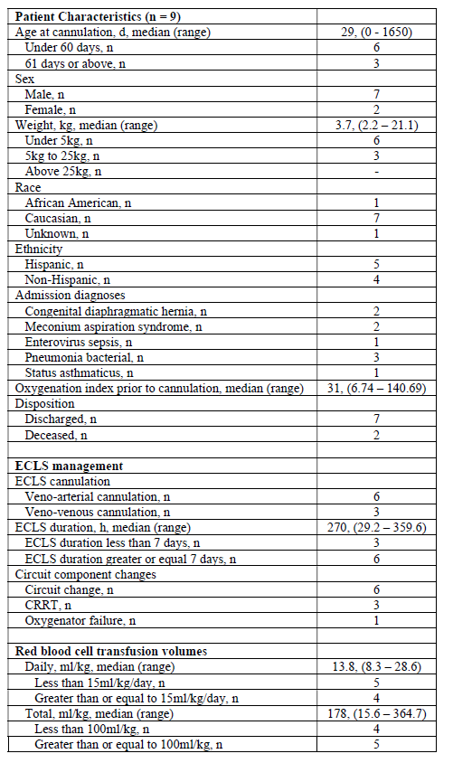
Results: Patient characteristics including ECLS management and transfusion practice are described in Table 1. All patients reported elevations in ferritin during ECLS, with 5 of 9 reporting elevations in serum iron after correction for age. Significant increases in ferritin were associated with veno-arterial cannulation (p = 0.026) and transfusion volume less than 100 ml/kg (p = 0.003). Iron elevation, however, was associated with higher daily (>15 ml/kg/day; p = 0.016) and cumulative transfusion volume (>100 mg/kg total; p = 0.013) as well as prolonged ECLS duration and patient age under 60 days of life. Plasma free hemoglobin elevation, consistent with acute hemolysis, did correlate with ferritin increase (p < 0.001). Three patients consented to post-decannulation MRI, with none demonstrating evidence of diffuse iron deposition despite elevated iron indices.Conclusion(s): Patient related factors including type of ECLS cannulation and volume of red blood cell transfusion may be associated with acute elevations in iron indices during ECLS. Despite acutely elevated iron indices, there was no evidence of increased iron deposition on MRI. Further investigation is warranted regarding the utility of iron storage indices during ECLS and patient related outcomes.
Ashley Sam CV - PAS.pdf
Summary of Patient Characteristics Including ECLS and Transfusion Exposures

