
Neonatal Infectious Diseases/Immunology
Category: Abstract Submission
Neonatal Infectious Diseases/Immunology: COVID-19
 photo")
Lillian Juttukonda, MD, PhD (she/her/hers)
Neonatology Fellow
Boston Children's Hospital
Boston, Massachusetts, United States
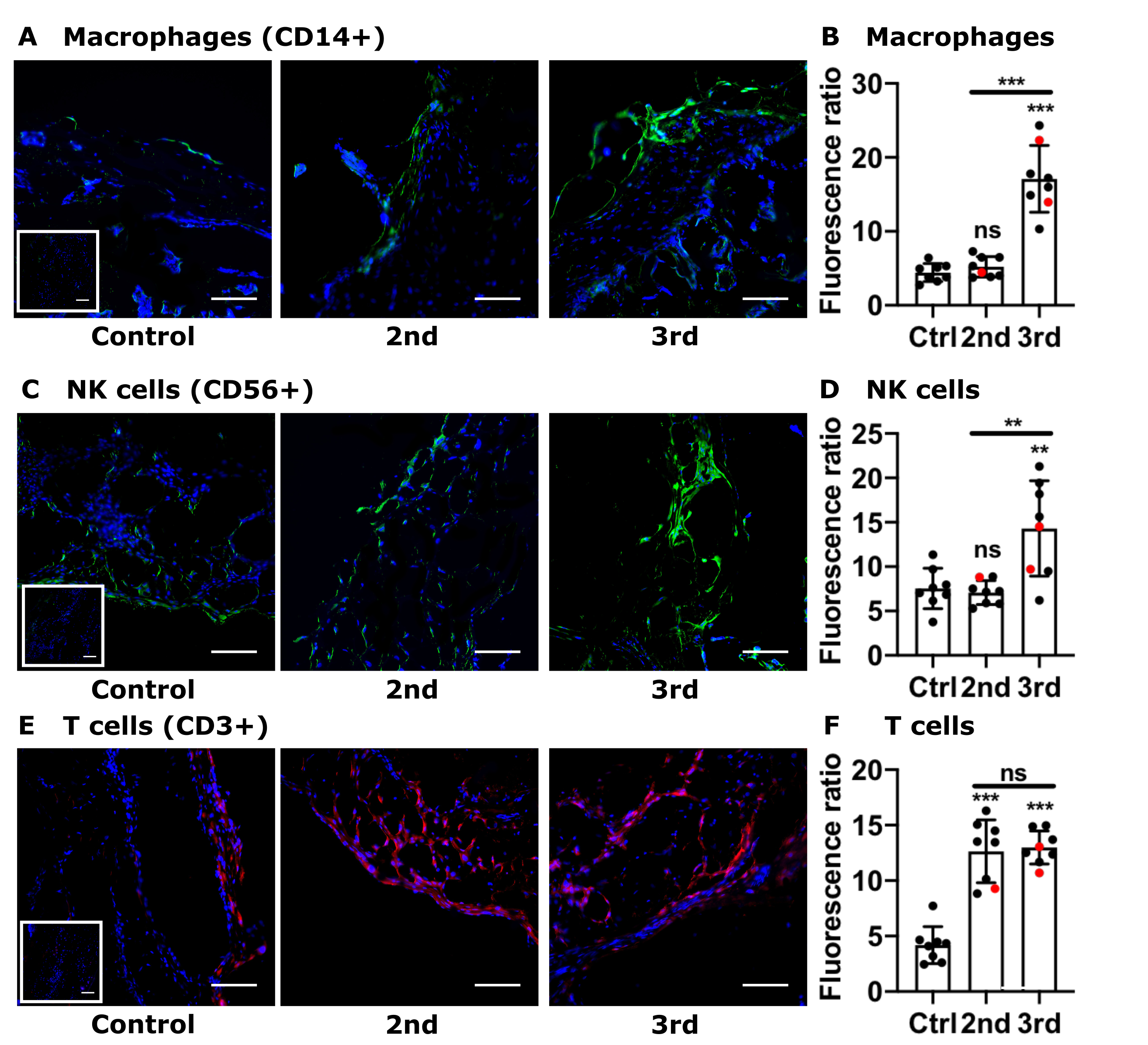 (A, C, E) Representative images (200x) of placental decidual areas stained for (A) CD14 (macrophage marker, green), (C) CD56 (NK cell marker, green), or (E) CD3 (T cell marker, red) immunofluorescence. (B, D, F) Graphical analysis of comparative fluorescence quantitation of (B) macrophages (CD14+), (E) NK cells (CD56+), or (F) T cells (CD3+). Red circles indicate placentas with positive staining for SARS-CoV-2 Spike protein. 2nd = 2nd Trimester COVID (n = 8); 3rd = 3rd Trimester COVID (n = 8); control/ctrl = negative for COVID (n = 8). ns = not significant; ** p < 0.01; ** p < 0.001 by ANOVA. Scale bar: 50µm; insets: secondary-only negative controls.
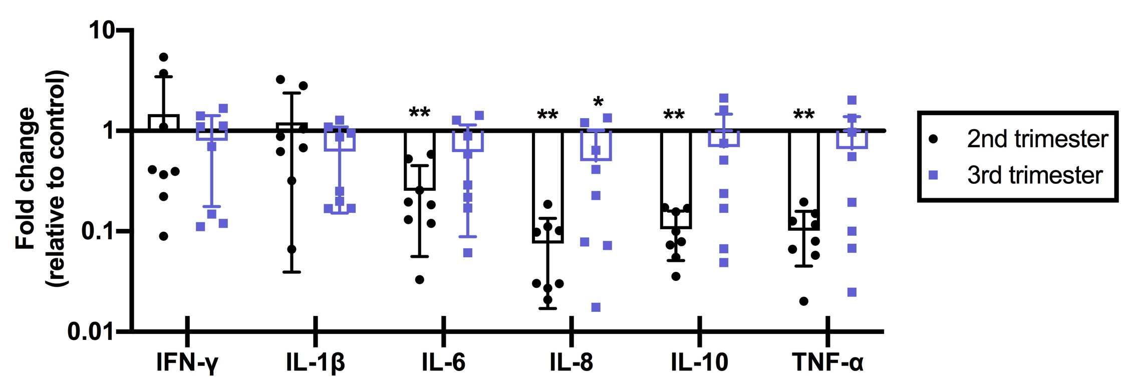 Fold-changes in cytokine abundance for IFN-, IL-1, IL-6, IL-8, IL-10, and TNF- by qRT-PCR of placental tissues from women with 2nd or 3nd trimester COVID infection (n = 8/group) calculated as fold-change compared to control (n = 8). ** p < 0.01; * p < 0.05.