Neonatal Neurology: Clinical
Category: Abstract Submission
Neurology 6: Neonatal Neurology Preterm Imaging
338 - Incidental findings on 3T brain MRI at term-equivalent age in a research cohort of very preterm infants and term-born controls
Monday, April 25, 2022
3:30 PM - 6:00 PM US MT
Poster Number: 338
Publication Number: 338.442
Publication Number: 338.442
Gemma Sullivan, University of Edinburgh, Edinburgh, Scotland, United Kingdom; Rory D. Teed, University of Edinburgh, Edinburgh, Scotland, United Kingdom; Alan J. Quigley, NHS Lothian, Edinburgh, Scotland, United Kingdom; Amy E. Corrigan, University of Edinburgh, Edinburgh, Scotland, United Kingdom; Kadi Vaher, University of Edinburgh, Edinburgh, Scotland, United Kingdom; Manuel Blesa, University of Edinburgh, Edinburgh, Scotland, United Kingdom; David Q. Stoye, National Health Service, St. Albans, England, United Kingdom; Jill Hall, University of Edinburgh, Edinburgh, Scotland, United Kingdom; Michael Thrippleton, University of Edinburgh, Edinburgh, Scotland, United Kingdom; Mark Bastin, University of Edinburgh, Edinburgh, Scotland, United Kingdom; James P. Boardman, University of Edinburgh, Edinburgh, Scotland, United Kingdom
- GS
Gemma Sullivan, MBChB PhD
Dr
University of Edinburgh
Edinburgh, Scotland, United Kingdom
Presenting Author(s)
Background: MRI is a valuable research tool to study the developing brain because it provides unprecedented information about morphology, structural/functional connectivity and microstructural properties of tissues. All neuroimaging research has the potential to detect incidental findings that can impact individuals and health services; knowledge of the types and prevalence of incidental findings in the neonatal population is growing.
Objective: To report incidental findings from 3T brain MRI in a research cohort of very preterm infants scanned at term-equivalent age and term-born controls.
Design/Methods: Participants were preterm infants born before 32 weeks’ gestation and healthy term-born controls recruited to the Theirworld Edinburgh Birth Cohort. Exclusion criteria were major congenital malformation, chromosomal abnormality, congenital infection and overt parenchymal brain injury. 3T MRI brain was performed during natural sleep at 38 to 44 weeks’ postmenstrual age. Images were reported by an experienced paediatric radiologist using a structured system and the type and frequency of incidental findings were recorded.
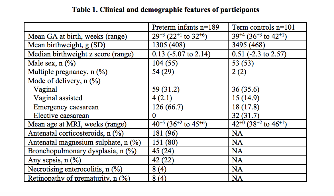
Results: Brain MRI data were available for 290 infants: 189 preterm infants and 101 term-born controls (Table 1). Incidental findings were detected in 12 (6%) preterm infants including developmental venous anomaly (n=2), focal ischaemic white matter injury (n=2), subarachnoid haemorrhage (n=2), giant cisterna magna (n=1), subdural collection following meningitis (n=1), unusually shaped choroid (n=1), suspected pontine cerebellar hypoplasia (n=1), arachnoid cyst (n=1) and a large middle cranial fossa cyst (n=1). Incidental findings were detected in 32 (32%) healthy term-born infants. The most frequent abnormality identified was subdural haemorrhage which affected 23 infants (23%). 4 had punctate white matter lesions, 3 had cysts identified (1 subependymal, 1 arachnoid, 1 neuroepithelial) and 4 had mild ventricular enlargement or prominence of extra-axial spaces. 1 infant had low-lying cerebellar tonsils and 1 had a developmental venous anomaly. Ten (6%) infants (8/189 preterm and 2/101 term) were referred to clinical services for further assessment with repeat MRI and/or multidisciplinary opinion.Conclusion(s): Incidental findings are relatively common in very preterm infants and term-born controls imaged at term-equivalent age and should be anticipated in research scans. These data will assist in optimising the accuracy of information sharing with families, informed consent processes and the development of care pathways for managing incidental findings.
Table 1. Clinical and demographic features of participants

