Neonatal Neurology: Translational
Category: Abstract Submission
Neurology 7: Neonatal Neurology Term Imaging
344 - Altered functional brain connectivity in HIV exposed neonates
Monday, April 25, 2022
3:30 PM - 6:00 PM US MT
Poster Number: 344
Publication Number: 344.443
Publication Number: 344.443
Nickie Andescavage, Children's National Health System, Washington, DC, United States; Josepheen D. Cruz, Children's National, Bethesda, MD, United States; Rachel K. Scott, MedStar Health Research Institute, Baltimore, MD, United States; Kushal Kapse, Developing Brain Institute, Washington, DC, United States; Catherine Lopez, Children's National Hospital, Washington, DC, United States; Haleema Saeed, MedStar Washington Hospital Center, Washington, DC, United States; Natella Y. Rakhmanina, George Washington University School of Medicine and Health Sciences, Washington, DC, United States; Catherine Limperopoulos, Children's National Health System, Washington, DC, United States; Nicole Andersen, Children's National Health System, Washington, DC, United States

Nickie Andescavage, MD
Associate Professor
Children's National Health System
Washington, District of Columbia, United States
Presenting Author(s)
Background: Despite conflicting data on neurocognitive outcomes in children exposed to HIV in utero, there remains significant concern for delayed cognitive and motor skills development in HIV-exposed uninfected (HEU) children compared to the HIV-unexposed and uninfected (HUU) children.
Objective: To compare emerging connectivity in the neonatal brain of HEU infants with HUU controls.
Design/Methods: We conducted a prospective, observational study of HEU and HUU control newborns. All infants were scanned without sedation using 3T GE scanner. We collected resting-state data using a T2-weighted gradient-echo planar imaging (EPI) sequence: echo/repetition time = 35/2000 ms, voxel size = 3.125 x 3.125 x 3 mm, flip angle = 60°, slice number = 29 to 40, number of volumes = 200 volumes representing 6 min 40 s of data. Preprocessing included temporal and spatial denoising. We defined regions of interest (ROIs) based on the neonatal Automatic Anatomic Labelling (AAL; 45 ROIs per hemisphere) and added manually refined cerebellar and brainstem labels for a total of 93 ROIs. We used the intrinsic connectivity distribution (ICD) to assess connectivity, computing the ICD for all voxels of the brain to explore regions that could serve as potential seeds and guide further investigation. We averaged normalized ICD values for all voxels within an ROI. Significant ROIs from ICD analysis were used to compare between-group connectivity in seed-based analysis.
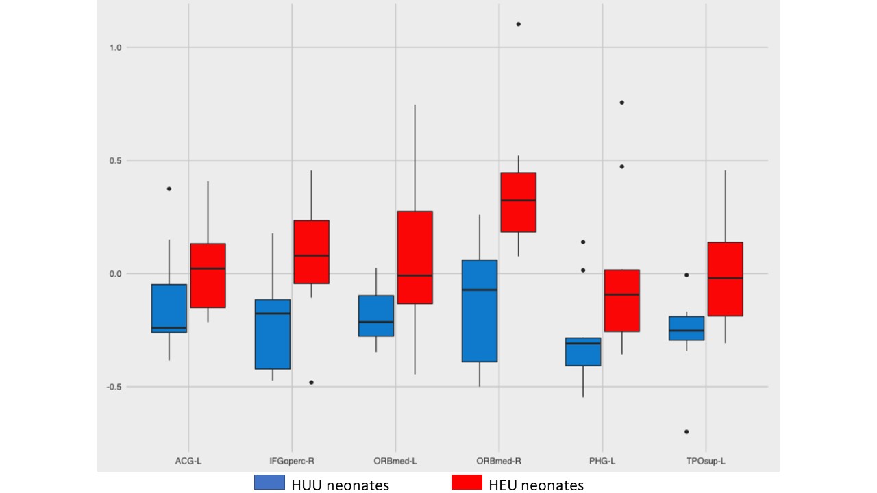
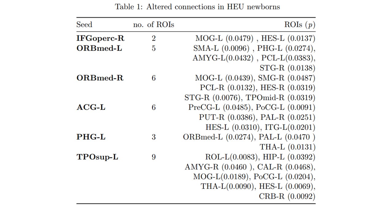
Results: We compared functional connectivity in 10 HEU newborns and 10 age matched HUU controls. Mean gestational age (GA) at birth: HUU 39.7±1.3 vs HEU 37.7±2.8; mean GA at MRI: HUU 42.7±1.8 vs HEU 42.6±1.9; 55% of neonates were male. All subjects had translational motion < 0.2mm and scan duration greater than 4 minutes (120 volumes). Six regions had significantly increased ICD for HEU newborns compared to controls (Figure 1). Seed-based analyses of these ROI’s identified 31 connections that were significantly altered in HEU newborns compared to HUU controls (Table 1).Conclusion(s): We report significant differences in emerging neural connectivity for HEU infants compared to HUU controls. These preliminary data may represent functional alterations in early neurodevelopment that may underlie delayed neurocognition in HEU children. Larger studies and long-term follow-up of the HEU children are warranted.
Figure 1 Functional connectivity differences measured using intrinsic connectivity distribution
Table 1 Altered connections in HEU newborns
Objective: To compare emerging connectivity in the neonatal brain of HEU infants with HUU controls.
Design/Methods: We conducted a prospective, observational study of HEU and HUU control newborns. All infants were scanned without sedation using 3T GE scanner. We collected resting-state data using a T2-weighted gradient-echo planar imaging (EPI) sequence: echo/repetition time = 35/2000 ms, voxel size = 3.125 x 3.125 x 3 mm, flip angle = 60°, slice number = 29 to 40, number of volumes = 200 volumes representing 6 min 40 s of data. Preprocessing included temporal and spatial denoising. We defined regions of interest (ROIs) based on the neonatal Automatic Anatomic Labelling (AAL; 45 ROIs per hemisphere) and added manually refined cerebellar and brainstem labels for a total of 93 ROIs. We used the intrinsic connectivity distribution (ICD) to assess connectivity, computing the ICD for all voxels of the brain to explore regions that could serve as potential seeds and guide further investigation. We averaged normalized ICD values for all voxels within an ROI. Significant ROIs from ICD analysis were used to compare between-group connectivity in seed-based analysis.
Results: We compared functional connectivity in 10 HEU newborns and 10 age matched HUU controls. Mean gestational age (GA) at birth: HUU 39.7±1.3 vs HEU 37.7±2.8; mean GA at MRI: HUU 42.7±1.8 vs HEU 42.6±1.9; 55% of neonates were male. All subjects had translational motion < 0.2mm and scan duration greater than 4 minutes (120 volumes). Six regions had significantly increased ICD for HEU newborns compared to controls (Figure 1). Seed-based analyses of these ROI’s identified 31 connections that were significantly altered in HEU newborns compared to HUU controls (Table 1).Conclusion(s): We report significant differences in emerging neural connectivity for HEU infants compared to HUU controls. These preliminary data may represent functional alterations in early neurodevelopment that may underlie delayed neurocognition in HEU children. Larger studies and long-term follow-up of the HEU children are warranted.
Figure 1
 Functional connectivity differences measured using intrinsic connectivity distribution
Table 1
 Altered connections in HEU newborns