Neonatal Fetal Nutrition & Metabolism
Category: Abstract Submission
Neonatal Fetal Nutrition & Metabolism I
277 - Impact of Nutritional Status on Total Brain Tissue Volumes of Preterm Infants
Friday, April 22, 2022
6:15 PM - 8:45 PM US MT
Poster Number: 277
Publication Number: 277.119
Publication Number: 277.119
Cyndi Valdes, University of Florida, Gainesville, FL, United States; Parvathi Nataraj, Mednax, Fort Myers, FL, United States; Katherine E. Kisilewicz, UF Health Shands Children's Hospital, Gainesville, FL, United States; Nikolay Bliznyuk, University of Florida, Gainesville, FL, United States; Livia Sura, University of Florida College of Medicine, Gainesville, FL, United States; Michael Weiss, University of Florida College of Medicine, Gainesville, FL, United States
.jpeg.jpg)
Cyndi Valdes, MD
PGY-5
University of Florida
Gainesville, Florida, United States
Presenting Author(s)
Background: The fetal brain is a highly metabolic organ and it consumes the greatest number of nutrient resources for its function and growth during the second and third trimester of gestation. For the very premature infant, this crucial period of brain development will occur in the ex-utero environment, where there is increased risk of poor growth and malnutrition due to their innate organ immaturity, underdeveloped nutrient absorption and cautious enteral feeding advancement. Poor brain growth in this critical period may result from inadequate early nutrition.
Objective: The purpose of this study is to evaluate if nutritional status is associated with brain growth utilizing recently published guidelines for malnutrition.
Design/Methods: The study was a retrospective chart review enrolling 64 infants born < 30 weeks’ gestation admitted to the NICU at UF Health between the years 2018 -2019. Exclusion criteria included death, incomplete nutritional data and intracranial anomalies. Thirty-three variables were obtained from the chart, including gestational age, anthropometrics, information on enteral nutrition and malnutrition scores. Brain MRIs obtained at term equivalent age (TEA) were analyzed using T2 weighted axial sequences with ITK-SNAP software to perform manual segmentation by 2 different investigators to ensure optimal brain volume reconstruction, excluding ventricles, brainstem and cerebellum. We compared the obtained brain volumes with the maximum change in weight and length over time, type of enteral feed, intracranial hemorrhages and gestational age at birth. Linear regression models were used and when feasible, these were reaffirmed using generalized additive models (GAMs) capable of capturing nonlinear associations between the response variable and the covariate(s).
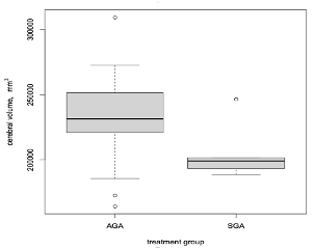
Results: Linear associations with brain volume at TEA and gestational age at birth and maximum change in weight over time were found (p < 0.05). Neonates that were small for gestational age (head sparing) had lower brain volumes at TEA compared to those that were appropriate for gestational age (p < 0.05).
Conclusion(s): With modeling using gestational age at birth and maximum changes in weight over time (Z- scores), infants tend to have a positive association with brain volumes at TEA. Further detailed analysis is ongoing to include other potential confounding variables to delineate the role of malnutrition on brain growth.
Resume - Cyndi ValdesRESUME Cyndi Paola Valdes Machorro 2022.pdf
Box plot demonstrating SGA infants having lower brain volumes compared to AGA

