
Neonatal Neurology: Clinical
Category: Abstract Submission
Neurology 6: Neonatal Neurology Preterm Imaging

Leah H. Fox, MD
Clinical Fellow
Cincinnati Children's Hospital Medical Center
CINCINNATI, Ohio, United States
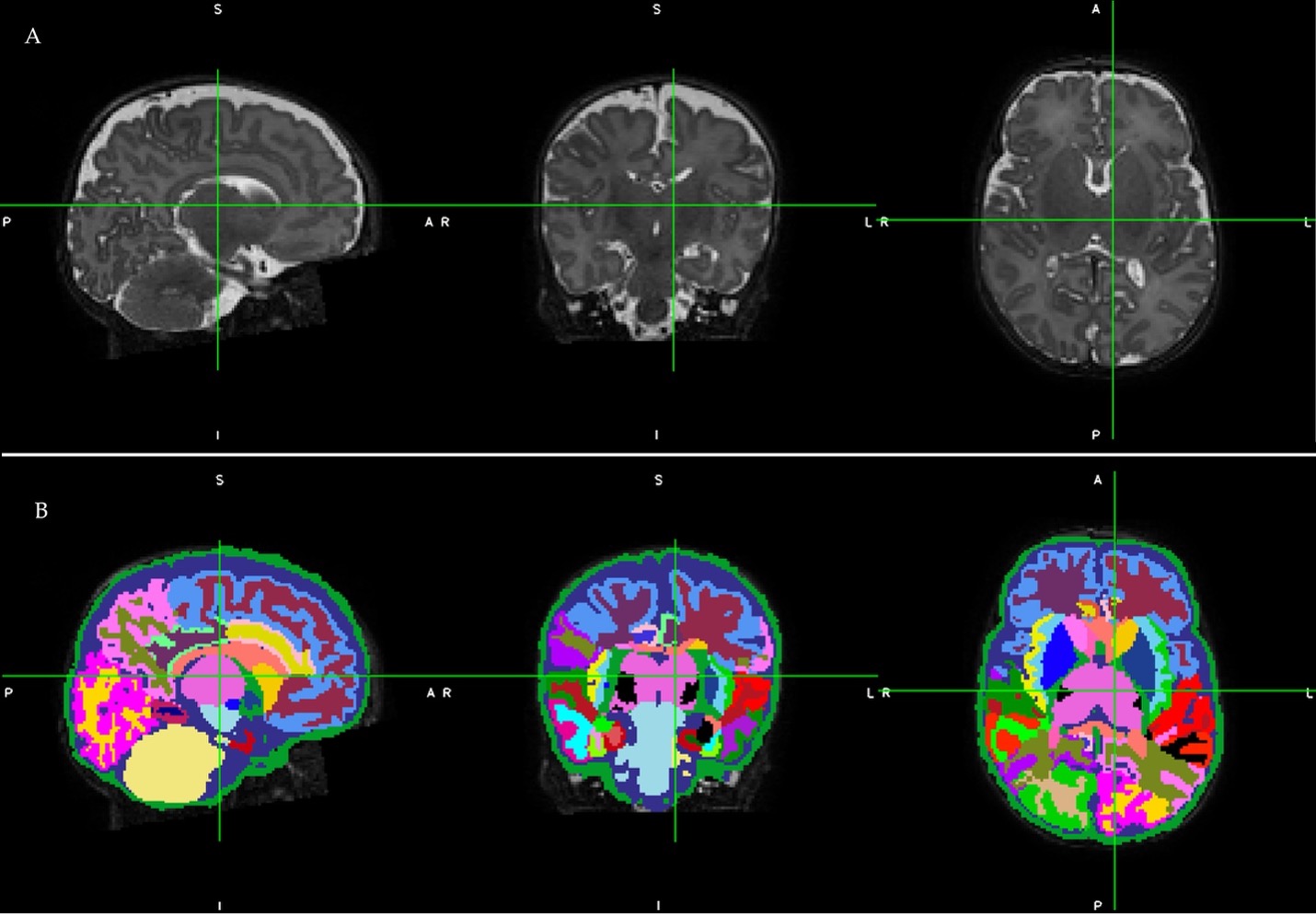 A representative participant’s raw T2-weighted midbrain MRI slices in sagittal, coronal, and axial orientations in panel A (top), and corresponding whole brain and regional cortical and tissue segmentations following automated processing using the Developing Human Connectome Project pipeline in panel B (bottom). Infant’s birth gestational age was 30.0 weeks and the MRI was performed at 41.1 weeks postmenstrual age. MRI Parameters: TE 166 ms, TR 18567 ms, FA 90°, voxel size 1.0×1.0× 1.0 mm3, and 3:43 minutes scan time.