Neonatal Neurology: Clinical
Category: Abstract Submission
Neurology 5: Neonatal Neurology Term Clinical
474 - Altered resting state functional connectivity in newborns with hypoxic ischemic encephalopathy assessed using high-density functional near-infrared spectroscopy
Sunday, April 24, 2022
3:30 PM - 6:00 PM US MT
Poster Number: 474
Publication Number: 474.343
Publication Number: 474.343
Lingkai Tang, Western University, London, ON, Canada; Lilian M. Kebaya, The University of Western Ontario - Schulich School of Medicine & Dentistry, London, ON, Canada; Talal Altamimi, The University of Western Ontario - Schulich School of Medicine & Dentistry, London, ON, Canada; Alexandra Kowalczyk, The University of Western Ontario - Schulich School of Medicine & Dentistry, London, ON, Canada; Melab S. Musabi, London Health Sciences Centre, London, ON, Canada; Sriya Roychaudhuri, The University of Western Ontario - Schulich School of Medicine & Dentistry, London, ON, Canada; Homa Vahidi, The University of Western Ontario - Schulich School of Medicine & Dentistry, London, ON, Canada; Paige Meyerink, LHSC, London, ON, Canada; Sandrine de Ribaupierre, The University of Western Ontario - Schulich School of Medicine & Dentistry, London, ON, Canada; Soume Bhattacharya, The University of Western Ontario - Schulich School of Medicine & Dentistry, London, ON, Canada; Keith St Lawrence, Lawson Health Research Institute, London, ON, Canada; Emma G. Duerden, Western University, London, ON, Canada

Lingkai Tang
PhD candidate
Western University
London, Ontario, Canada
Presenting Author(s)
Background: Hypoxic-Ischemic Encephalopathy (HIE) is a brain injury resulting from lack of oxygen to the brain during the antenatal period and has a prevalence of 1.5 cases per 1000 live births. HIE can lead to premature mortality, as well as a variety of acute and long-term morbidities. Therapeutic hypothermia (TH) is the standard of care for infants with HIE and has neuroprotective effects; yet, the mechanisms remain unknown. Functional near-infrared spectroscopy (fNIRS) is a non-invasive, portable, brain scanning tool that measures changes of cerebral oxygenation through determining concentrations of oxygenated and deoxygenated hemoglobin (HbO and Hbr). High-density fNIRS offers the ability to assess functional connectivity, a key marker of brain health in neonates with brain injury. Better understanding of the neural networks influenced by TH may aid in predicting functional outcomes in HIE.
Objective: To determine whether the patterns of resting state functional connectivity (RSFC) in term-born neonates with HIE in the post-cooling period, differ compared to healthy newborns, using high-density fNIRS.
Design/Methods: 18 neonates with HIE (median postmenstrual age [PMA]=38.8) and 17 term born healthy newborns (PMA=39.1) were recruited from the London Health Sciences Centre. Newborns with HIE underwent TH during the first week of life. Resting-state fNIRS data (6 minutes) were collected in newborns with HIE at the bedside in the post-cooling period. In healthy newborns, fNIRS data were collected within the first 8 hours of life. Preprocessing included motion correction, band-pass filtering and application of modified Beer-Lambert law. RSFC was calculated as the correlation coefficients amongst the time courses of the fNIRS channels with HbO and Hbr changes. Measuring local RSFC based on graph theory, the degree centrality ([DC] i.e., sum of connectivity) was calculated for each channel. T-tests were applied to the normalized DC values to determine significant group differences.
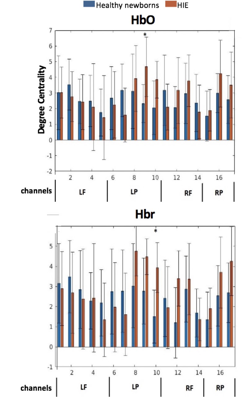
Results: Term-born neonates with HIE had significantly increased functional connectivity, reflected in the increased DC values, in the left parietal lobe channels for both HbO (p=0.0204) and Hbr (p=0.0034, Fig 1) resting-state networks. Conclusion(s): TH is associated with regionally-specific increases in functional connectivity patterns in newborns with HIE, which may be reflective of ongoing alterations in cerebral hemodynamics as a result of perinatal hypoxia-ischemia. Results highlight the importance of using bedside measurements of cerebral hemodynamics to better characterize brain health in newborns with HIE.
Degree centrality of fNIRS channels Fig. 1. Degree centrality (y-axis) of HbO and Hbr channels (x-axis) reflecting the sum of connectivity to each channel. Abbreviations, LF, left frontal, LP, left parietal; RF: right frontal; RP: right parietal. * p-value < 0.05, false discovery rate corrected.
Objective: To determine whether the patterns of resting state functional connectivity (RSFC) in term-born neonates with HIE in the post-cooling period, differ compared to healthy newborns, using high-density fNIRS.
Design/Methods: 18 neonates with HIE (median postmenstrual age [PMA]=38.8) and 17 term born healthy newborns (PMA=39.1) were recruited from the London Health Sciences Centre. Newborns with HIE underwent TH during the first week of life. Resting-state fNIRS data (6 minutes) were collected in newborns with HIE at the bedside in the post-cooling period. In healthy newborns, fNIRS data were collected within the first 8 hours of life. Preprocessing included motion correction, band-pass filtering and application of modified Beer-Lambert law. RSFC was calculated as the correlation coefficients amongst the time courses of the fNIRS channels with HbO and Hbr changes. Measuring local RSFC based on graph theory, the degree centrality ([DC] i.e., sum of connectivity) was calculated for each channel. T-tests were applied to the normalized DC values to determine significant group differences.
Results: Term-born neonates with HIE had significantly increased functional connectivity, reflected in the increased DC values, in the left parietal lobe channels for both HbO (p=0.0204) and Hbr (p=0.0034, Fig 1) resting-state networks. Conclusion(s): TH is associated with regionally-specific increases in functional connectivity patterns in newborns with HIE, which may be reflective of ongoing alterations in cerebral hemodynamics as a result of perinatal hypoxia-ischemia. Results highlight the importance of using bedside measurements of cerebral hemodynamics to better characterize brain health in newborns with HIE.
Degree centrality of fNIRS channels
 Fig. 1. Degree centrality (y-axis) of HbO and Hbr channels (x-axis) reflecting the sum of connectivity to each channel. Abbreviations, LF, left frontal, LP, left parietal; RF: right frontal; RP: right parietal. * p-value < 0.05, false discovery rate corrected.