Neonatal Clinical Trials
Category: Abstract Submission
Neonatal Clinical Trials I
429 - Role of point of care ultrasonography for endotracheal tube placement in neonates: A randomized controlled study
Saturday, April 23, 2022
3:30 PM - 6:00 PM US MT
Poster Number: 429
Publication Number: 429.223
Publication Number: 429.223
pranaya mall, ABVIMS & Dr.RML HOSPITAL NEW DELHI, new delhi, Delhi, India; Arti Maria, India, Delhi, Delhi, India; yashvant singh, Dr RML Hospital New Delhi, New Delhi, Delhi, India
- pm
pranaya mall
ABVIMS & Dr.RML HOSPITAL NEW DELHI
new delhi, Delhi, India
Presenting Author(s)
Background: Endotracheal intubation is a common procedure in the management of respiratory distress in critically ill neonates. However, optimal positioning of endotracheal tube tip (ETT) during both elective as well as emergency conditions is challenging. At present radiographic confirmation is the gold standard to identify the correct ETT placement but it often takes time and is also associated with the risk of radiation exposure. Hence, this study was conducted to evaluate the role of ultrasonography (USG) for optimal placement of ET tube confirmed by chest X- ray in babies requiring endotracheal intubation.
Objective: To compare the malposition rates of the ETT in the “Tochen's plus ultrasonography method” as compared to " Tochen’s method", confirmed in X-ray chest among neonates requiring mechanical ventilation.
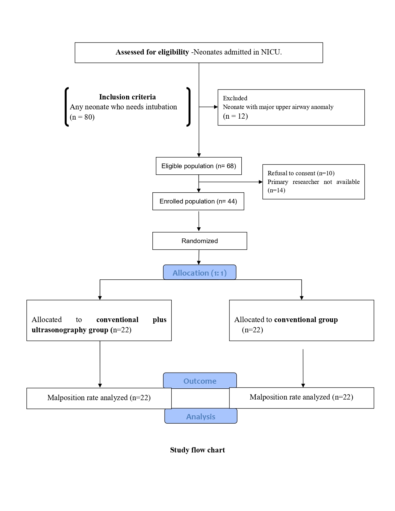
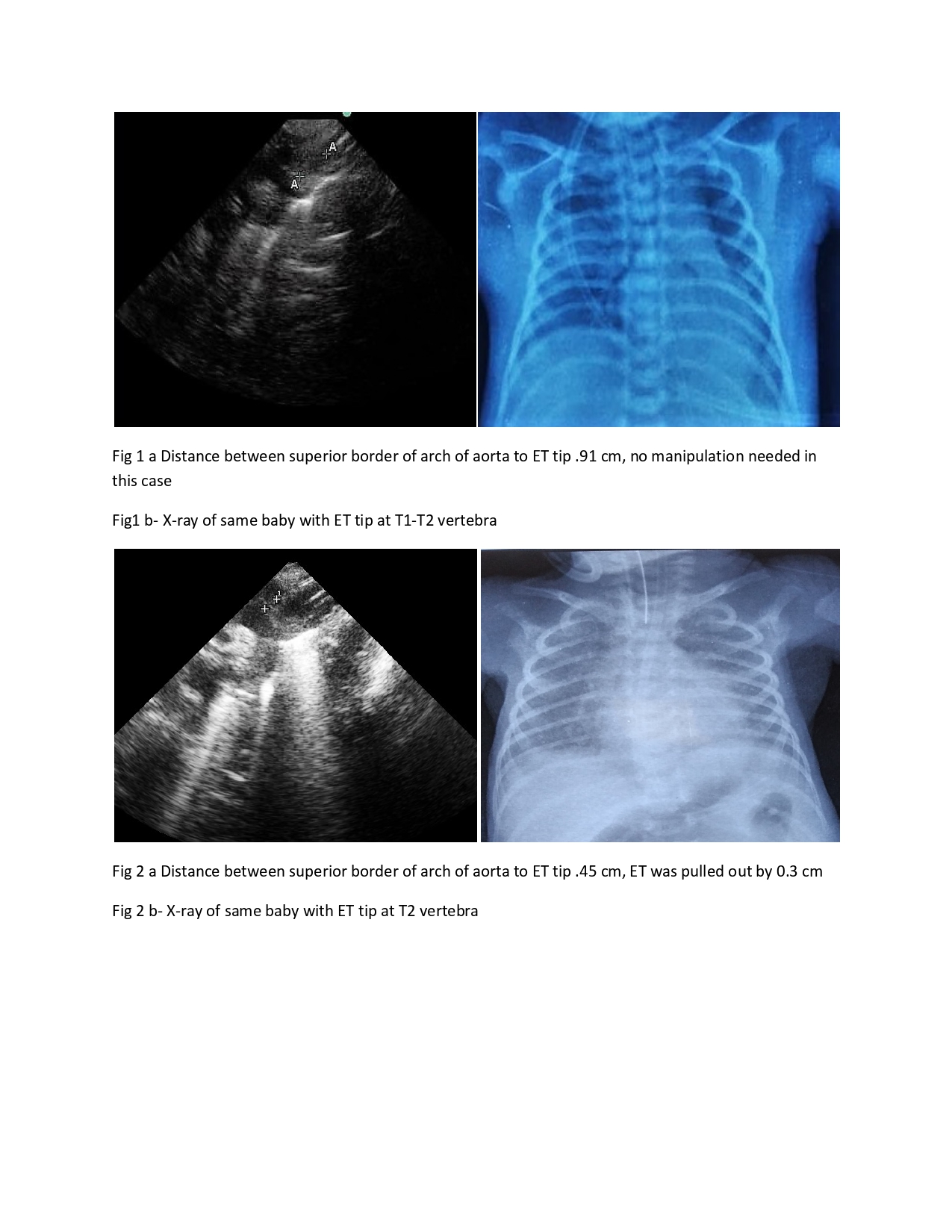
Design/Methods: Design- Open label Randomized controlled study with parallel-group.Settings-Neonatal intensive care unit of a tertiary care hospital. Participants- Forty-four neonates selected by computer-generated random number table and allocated into two groups with equal allocation (1:1) ratio. Intervention- Initially, the depth of insertion of ETT was decided based on Tochen’s formula (weight (in kg) +6 cm). Thereafter, the USG chest was done and ETT was manipulated to an optimal position (i.e. 0.5-1 cm from the superior border of arch of aorta). Final positioning of the ETT tube was based on radiographic confirmation (T1-T2 vertebra on X-ray chest). Control- Initially, the depth of insertion of ETT was decided based on Tochen’s formula. Final positioning of the ETT tube was based on radiographic confirmation.Primary outcome- To compare the malposition rates among intervention and control group.
Results: The malposition rate in intervention group (3/22) was significantly lower as compared to the control group (14/22) (RR 0.21, CI 0.07-0.64, P-value < .001). Acute complication rates related to malposition in intervention group (4/22) were no different from control (5/22) (RR 0.8, CI 0.24-2.5, P-value >0.5).Conclusion(s): Use of USG to complement the Tochen’s formula significantly reduces malposition rates compared to Tochen’s formula alone, measured by gold standard of X-ray chest.
Study flow chart
Endotracheal tube tip position in USG and X-rays
Objective: To compare the malposition rates of the ETT in the “Tochen's plus ultrasonography method” as compared to " Tochen’s method", confirmed in X-ray chest among neonates requiring mechanical ventilation.
Design/Methods: Design- Open label Randomized controlled study with parallel-group.Settings-Neonatal intensive care unit of a tertiary care hospital. Participants- Forty-four neonates selected by computer-generated random number table and allocated into two groups with equal allocation (1:1) ratio. Intervention- Initially, the depth of insertion of ETT was decided based on Tochen’s formula (weight (in kg) +6 cm). Thereafter, the USG chest was done and ETT was manipulated to an optimal position (i.e. 0.5-1 cm from the superior border of arch of aorta). Final positioning of the ETT tube was based on radiographic confirmation (T1-T2 vertebra on X-ray chest). Control- Initially, the depth of insertion of ETT was decided based on Tochen’s formula. Final positioning of the ETT tube was based on radiographic confirmation.Primary outcome- To compare the malposition rates among intervention and control group.
Results: The malposition rate in intervention group (3/22) was significantly lower as compared to the control group (14/22) (RR 0.21, CI 0.07-0.64, P-value < .001). Acute complication rates related to malposition in intervention group (4/22) were no different from control (5/22) (RR 0.8, CI 0.24-2.5, P-value >0.5).Conclusion(s): Use of USG to complement the Tochen’s formula significantly reduces malposition rates compared to Tochen’s formula alone, measured by gold standard of X-ray chest.
Study flow chart

Endotracheal tube tip position in USG and X-rays

